06.04.11 – Congenital anomaly of left-sided atrioventricular valve in double inlet ventricle

IPCCC Term CONGENITAL ANOMALY OF LEFT-SIDED ATRIOVENTRICULAR VALVE IN DOUBLE INLET VENTRICLE

IPCCC Code 06.04.11

ICD-11 Code PENDING

Synonyms PENDING

Abbreviations PENDING

IPCCC Definition A congenital cardiac malformation in association with double inlet ventricle (excluding common atrioventricular valve) in which the atrioventricular valve morphology cannot be determined. This term should be used for the left-sided atrioventricular valve in those hearts.

[restabs alignment=”osc-tabs-right” pills=”nav-pills” responsive=”true” icon=”true” text=”More”][restab title=”Anatomic Specimen” active=”active”]

06-04-11 
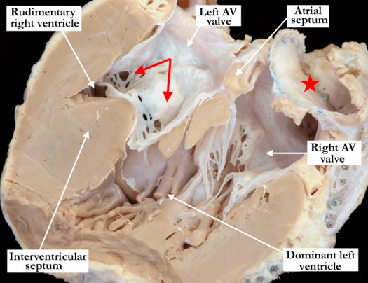
This heart is viewed from posterior to anterior and has been cut in a four chamber echocardiographic plane. There is a double inlet left ventricle with an abnormality of the left-sided atrioventricular valve. It straddles the interventricular septum with the tension apparatus extending across the ventricular septal defect into the rudimentary, incomplete right ventricle. This heart was explanted following a Fontan procedure. Note the remnant of the right-sided atrial baffle (red star).

[/restab][restab title=”Echocardiogram”]Content for Echocardiogram[/restab][restab title=”Computerized Axial Tomography”]Content for Computerized Axial Tomography[/restab][restab title=”MRI”]MRI[/restab][restab title=”Angiography”]Content for Angiography[/restab][restab title=”Intraoperative Videos”]Content for Intraoperative Videos[/restab][restab title=”Other”]Other[/restab][/restabs]

05.06.02 – Common atrium with separate atrioventricular junctions

IPCCC Term COMMON ATRIUM WITH SEPARATE ATRIOVENTRICULAR JUNCTIONS

IPCCC Code 05.06.02

ICD-11 Code PENDING

Synonyms PENDING

Abbreviations PENDING

IPCCC Definition A congenital cardiovascular malformation in which there is near-complete absence of the interatrial septum.

[restabs alignment=”osc-tabs-right” pills=”nav-pills” responsive=”true” icon=”true” text=”More”][restab title=”Anatomic Specimen” active=”active”]

05-06-02 In this postero-inferior view, the inferior aspects of both the right and left atrial chambers have been dissected and lifted away from the base of the heart. The majority of the atrial septum is absent in this heart with separate atrioventricular connections. There is a small portion of the interatrial septum (red dots) remaining near the atrioventricular junctions, rendering this as a common atrium. There are still two atrial chambers.

[/restab][restab title=”Echocardiogram”]Content for Echocardiogram[/restab][restab title=”Computerized Axial Tomography”]Content for Computerized Axial Tomography[/restab][restab title=”MRI”]MRI[/restab][restab title=”Angiography”]Content for Angiography[/restab][restab title=”Intraoperative Videos”]Content for Intraoperative Videos[/restab][restab title=”Other”]Other[/restab][/restabs]

09.43.13 – Single coronary supplying all of heart

IPCCC Term SINGLE CORONARY SUPPLYING ALL OF HEART

IPCCC Code 09.43.13

ICD-11 Code PENDING

Synonyms PENDING

Abbreviations PENDING

IPCCC Definition A congenital cardiovascular malformation in which a solitary coronary artery supplies the myocardium.

[restabs alignment=”osc-tabs-right” pills=”nav-pills” responsive=”true” icon=”true” text=”More”][restab title=”Anatomic Specimen” active=”active”]

09-43-13 In this left anterior lateral view the pulmonary trunk is folded forward and the subpulmonary muscular infundibulum has been dissected. The red dots mark the junction of the pulmonary arterial wall with the infundibular muscle. There is a single coronary artery with the left main coronary artery arising from the proximal right coronary artery and extending between the arterial trunks within the infundibular muscle. As it emerges from the muscle to the left of the pulmonary trunk, the artery divided in the usual fashion to the anterior interventricular and circumflex branches. The right coronary artery took its usual course within the right atrioventricular groove.

[/restab][restab title=”Echocardiogram”]Content for Echocardiogram[/restab][restab title=”Computerized Axial Tomography”]Content for Computerized Axial Tomography[/restab][restab title=”MRI”]MRI[/restab][restab title=”Angiography”]Content for Angiography[/restab][restab title=”Intraoperative Videos”]Content for Intraoperative Videos[/restab][restab title=”Other”]Other[/restab][/restabs]

09.10.71 – Congenital pulmonary arterial (branch) hypoplasia

IPCCC Term CONGENITAL PULMONARY ARTERIAL (BRANCH) HYPOPLASIA

IPCCC Code 09.10.71

ICD-11 Code PENDING

Synonyms PENDING

Abbreviations PENDING

IPCCC Definition A congenital cardiovascular malformation in which there is diffuse luminal narrowing of a pulmonary arterial branch (below the lower limit of normal adjusted for body size).

[restabs alignment=”osc-tabs-right” pills=”nav-pills” responsive=”true” icon=”true” text=”More”][restab title=”Anatomic Specimen” active=”active”]

[su_carousel source=”media: 6258,6259″ limit=”27″ width=”460″ height=”510″ responsive=”no” items=”1″ title=”no” centered=”no” mousewheel=”no” autoplay=”0″] The right and left pulmonary arteries in this heart are hypoplastic bilaterally and along their entire length. Image #1 is a left lateral view showing the pulmonary trunk, the hypoplastic left pulmonary artery, the arterial duct (black star) and the aortic arch. The hypoplastic left pulmonary arterial wall is very thin, and it is stenotic along its entire length. In Image #2, the arterial trunks are folded anteriorly over the ventricular mass, showing the uniform hypoplasia of both the right and left pulmonary arteries as they extend to the hilar regions of the lungs.

[/restab][restab title=”Echocardiogram”]Content for Echocardiogram.[/restab][restab title=”Computerized Axial Tomography”]Content for Computerized Axial Tomography.[/restab][restab title=”Angiography”]Content for Angiography.[/restab][restab title=”Intraoperative Videos”]Content for Intraoperative Videos.[/restab][/restabs]

01.05.01 + 01.04.00 + 07.10.00 + 07.09.01 – Transposition of the great arteries with concordant atrioventricular connections and ventricular septal defect and lev

IPCCC Term TRANSPOSITION OF THE GREAT ARTERIES WITH CONCORDANT ATRIOVENTRICULAR CONNECTIONS AND VENTRICULAR SEPTAL DEFECT AND LEFT VENTRICULAR OUTFLOW TRACT OBSTRUCTION

IPCCC Code 01.05.01 + 01.04.00 + 07.10.00 + 07.09.01

ICD-11 Code PENDING

Synonyms PENDING

Abbreviations PENDING

IPCCC Definition A congenital cardiovascular malformation in which the morphologically right atrium connects to the morphologically right ventricle, the morphologically left atrium connects to the morphologically left ventricle, the morphologically right ventricle connects to the aorta, the morphologically left ventricle connects to the pulmonary trunk, one or more ventricular septal defects are present, and left ventricular outflow tract obstruction is present.

[restabs alignment=”osc-tabs-right” pills=”nav-pills” responsive=”true” icon=”true” text=”More”][restab title=”Anatomic Specimen” active=”active”]

[su_carousel source=”media: 6263,6264″ limit=”27″ width=”460″ height=”510″ responsive=”no” items=”1″ title=”no” centered=”no” mousewheel=”no” autoplay=”0″] In Image #1, the anterior free wall of the right ventricle and the aorta have been removed. This heart has concordant atrioventricular connections with discordant ventriculo-arterial connections along with a ventricular septal defect and posterior deviation of the outlet septum. The ventricular septal defect is within the outlet component of the right ventricle and extends to become perimembranous with malalignment of the outlet septum. The tendinous cords that support the tricuspid valve extend across the inferior aspect of the defect. Image #2 shows the left ventricular outflow tract from the same heart as in the left panel. The malalignment of the outlet septum narrows the left ventricular outflow tract and causes significant pulmonary stenosis. The pulmonary valve is bicuspid.

[/restab][restab title=”Echocardiogram”]Content for Echocardiogram.[/restab][restab title=”Computerized Axial Tomography”]Content for Computerized Axial Tomography.[/restab][restab title=”Angiography”]Content for Angiography.[/restab][restab title=”Intraoperative Videos”]Content for Intraoperative Videos.[/restab][/restabs]

09.01.19 – Common arterial trunk (truncus arteriosus) with pulmonary dominance and aortic coarctation

IPCCC Term COMMON ARTERIAL TRUNK (TRUNCUS ARTERIOSUS) WITH PULMONARY DOMINANCE AND AORTIC COARCTATION

IPCCC Code 09.01.19

ICD-11 Code PENDING

Synonyms PENDING

Abbreviations PENDING

IPCCC Definition A congenital cardiovascular malformation in which a common arterial trunk is associated with aortic coarctation.

[restabs alignment=”osc-tabs-right” pills=”nav-pills” responsive=”true” icon=”true” text=”More”][restab title=”Anatomic Specimen” active=”active”]

[su_carousel source=”media: 6239,6240″ limit=”27″ width=”460″ height=”510″ responsive=”no” items=”1″ title=”no” centered=”no” mousewheel=”no” autoplay=”0″] Image #1 is an anterior view illustrating the outward appearance of a pulmonary dominant common arterial trunk with an associated tubular narrowing or coarctation of the aortic arch between the left carotid and left subclavian arteries. Note the large arterial duct between the pulmonary component of the common trunk and the descending aorta. In Image #2 the common arterial trunk has been opened and is viewed anteriorly. The pulmonary component is dominant with the small aortic component marked with the black dots. The truncal valve consists of three leaflets and there is a muscular, subarterial ventricular septal defect (interventricular communication) with a postero-inferior muscular bar (caudal arm of the septomarginal trabeculation) separating the arterial from the atrioventricular valve. (yellow Y – septomarginal trabeculation)

[/restab][restab title=”Echocardiogram”]Content for Echocardiogram.[/restab][restab title=”Computerized Axial Tomography”]Content for Computerized Axial Tomography.[/restab][restab title=”Angiography”]Content for Angiography.[/restab][restab title=”Intraoperative Videos”]Content for Intraoperative Videos.[/restab][/restabs]

09.02.18 – Congenital truncal valvar stenosis

IPCCC Term CONGENITAL TRUNCAL VALVAR STENOSIS

IPCCC Code 09.02.18

ICD-11 Code PENDING

Synonyms PENDING

Abbreviations PENDING

IPCCC Definition A congenital cardiovascular malformation in which there is obstruction to flow through the truncal valve due to narrowing or stricture.

[restabs alignment=”osc-tabs-right” pills=”nav-pills” responsive=”true” icon=”true” text=”More”][restab title=”Anatomic Specimen” active=”active”]

09-02-18 In this anterior view the right ventricular outflow tract is opened to show the arterial valve in this heart with a common arterial trunk. The valve is quadricuspid, thickened and dysplastic. There is valvar stenosis with post-stenotic dilation of the common trunk. The pulmonary component branches very low from the common trunk, at the sinutubular junction and extending into the adjacent sinus of Valsalva. There is a restrictive, subarterial, muscular ventricular septal defect.

[/restab][restab title=”Echocardiogram”]Content for Echocardiogram[/restab][restab title=”Computerized Axial Tomography”]Content for Computerized Axial Tomography[/restab][restab title=”MRI”]MRI[/restab][restab title=”Angiography”]Content for Angiography[/restab][restab title=”Intraoperative Videos”]Content for Intraoperative Videos[/restab][restab title=”Other”]Other[/restab][/restabs]

09.41.03 – Anomalous origin of left coronary artery from pulmonary artery

IPCCC Term ANOMALOUS ORIGIN OF LEFT CORONARY ARTERY FROM PULMONARY ARTERY

IPCCC Code 09.41.03

ICD-11 Code PENDING

Synonyms PENDING

Abbreviations PENDING

IPCCC Definition A congenital cardiovascular malformation in which a coronary artery originates from the pulmonary trunk or one of its branches.

[restabs alignment=”osc-tabs-right” pills=”nav-pills” responsive=”true” icon=”true” text=”More”][restab title=”Anatomic Specimen” active=”active”]

[su_carousel source=”media: 6244,6245″ limit=”27″ width=”460″ height=”510″ responsive=”no” items=”1″ title=”no” centered=”no” mousewheel=”no” autoplay=”0″] The images are from the same heart, the left-hand image is viewed anteriorly with the pulmonary valve, pulmonary trunk and both right and left pulmonary arteries opened. The left coronary orifice arises anomalously from the proximal right pulmonary artery. In the right-hand image the left pulmonary artery is retracted anteriorly to show the origin of the left main coronary artery from the right pulmonary artery. The left main coronary artery has an extremely long course prior to reaching the epicardial surface where it then bifurcates in the usual fashion to the anterior interventricular and circumflex branches.

[/restab][restab title=”Echocardiogram”]Content for Echocardiogram.[/restab][restab title=”Computerized Axial Tomography”]Content for Computerized Axial Tomography.[/restab][restab title=”Angiography”]Content for Angiography.[/restab][restab title=”Intraoperative Videos”]Content for Intraoperative Videos.[/restab][/restabs]

09.18.01 – Aneurysm of aortic sinus of Valsalva

IPCCC Term ANEURYSM OF AORTIC SINUS OF VALSALVA

IPCCC Code 09.18.01

ICD-11 Code PENDING

Synonyms PENDING

Abbreviations PENDING

IPCCC Definition A congenital cardiovascular malformation in which there is dilation of a single aortic sinus of Valsalva.

[restabs alignment=”osc-tabs-right” pills=”nav-pills” responsive=”true” icon=”true” text=”More”][restab title=”Anatomic Specimen” active=”active”]

[su_carousel source=”media: 4982,4983″ limit=”27″ width=”460″ height=”510″ responsive=”no” items=”1″ title=”no” centered=”no” mousewheel=”no” autoplay=”0″] Image #1 is a close up, superior view of an opened aortic valve. The right coronary aortic valvar leaflet is retracted with a suture to demonstrate the dilated sinus of Valsalva. The aortic root in this case was poorly supported with a large membranous septum and no muscular support beneath the right coronary leaflet. Only the left coronary leaflet was supported by muscle (not shown). Note the left coronary orifice with its origin near the attachment of the left coronary leaflet at the sinutubular junction. In Image #2, the right atrioventricular junction is viewed anteriorly and in anatomic position. The aneurysm at the atrioventricular junction has been transected. The sectioned atrial myocardium and tricuspid valve demonstrate the close proximity of the aorta, atrioventricular and interventricular components of the membranous septum. The sinus of Valsalva aneurysm caused an ovoid, saccular dilation of the tricuspid valve hinge point that eventually perforated.

[/restab][restab title=”Echocardiogram”]Content for Echocardiogram.[/restab][restab title=”Computerized Axial Tomography”]Content for Computerized Axial Tomography.[/restab][restab title=”Angiography”]Content for Angiography.[/restab][restab title=”Intraoperative Videos”]Content for Intraoperative Videos.[/restab][/restabs]

07.12.08 – Doubly committed juxta-arterial outlet ventricular septal defect with posteriorly malaligned outlet septum

IPCCC Term DOUBLY COMMITTED JUXTA-ARTERIAL OUTLET VENTRICULAR SEPTAL DEFECT WITH POSTERIORLY MALALIGNED OUTLET SEPTUM

IPCCC Code 07.12.08

ICD-11 Code PENDING

Synonyms PENDING

Abbreviations PENDING

IPCCC Definition A congenital cardiac malformation in which there is a ventricular septal defect that opens to the outlet of the right ventricle, with a muscular postero-inferior border when viewed from the right ventricle, and in which the fibrous outlet septum, or the area of continuity between the aortic and pulmonary valves, is malaligned in postero-caudal fashion relative to the apical part of the muscular septum such that there is obstruction to the left ventricular outflow tract.

[restabs alignment=”osc-tabs-right” pills=”nav-pills” responsive=”true” icon=”true” text=”More”][restab title=”Anatomic Specimen” active=”active”]

[su_carousel source=”media: 6255,6256″ limit=”27″ width=”460″ height=”510″ responsive=”no” items=”1″ title=”no” centered=”no” mousewheel=”no” autoplay=”0″] In Image #1, the pulmonary outflow is viewed anteriorly, in anatomic position. There is a ventricular septal defect that opens to the outlet of the right ventricle, with a muscular postero-inferior border (red dots) when viewed from the right ventricle. The area of fibrous continuity between the aortic and pulmonary valves is malaligned in postero-caudal fashion along with the adjacent muscular portion of the outlet septum, such that there is obstruction to the left ventricular outflow tract. In Image #2 the left ventricular outflow of the same heart shown in the left hand image demonstrates the left ventricular outflow tract viewed posteriorly and in anatomic position. The muscular component of the outlet septum joins the area of fibrous continuity between the pulmonary and aortic valves and is deviated in a postero-caudal direction causing left ventricular outflow tract obstruction.

[/restab][restab title=”Echocardiogram”]Content for Echocardiogram.[/restab][restab title=”Computerized Axial Tomography”]Content for Computerized Axial Tomography.[/restab][restab title=”Angiography”]Content for Angiography.[/restab][restab title=”Intraoperative Videos”]Content for Intraoperative Videos.[/restab][/restabs]