07.10.01 – Central perimembranous ventricular septal defect

IPCCC Term CENTRAL PERIMEMBRANOUS VENTRICULAR SEPTAL DEFECT

IPCCC Code 07.10.01

ICD-11 Code PENDING

Synonyms PENDING

Abbreviations PENDING

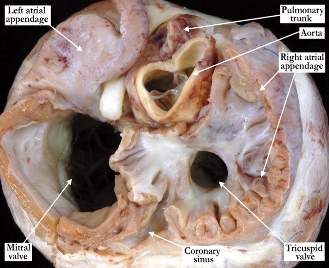
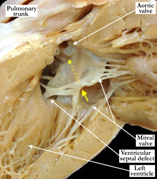
IPCCC Definition A congenital cardiovascular malformation in which there is a ventricular septal defect that 1) occupies the space that is usually closed by the interventricular part of the membranous septum, 2) is adjacent to the area of fibrous continuity between the leaflets of an atrioventricular valve and an arterial valve, and 3) is located at the center of the base of the ventricular mass.

[restabs alignment=”osc-tabs-right” pills=”nav-pills” responsive=”true” icon=”true” text=”More”][restab title=”Anatomic Specimen” active=”active”]

[su_carousel source=”media: 6253,6254″ limit=”27″ width=”460″ height=”510″ responsive=”no” items=”1″ title=”no” centered=”no” mousewheel=”no” autoplay=”0″] In the first image, the central ventricular septal defect lies at the apex of the triangle of Koch and the supraventricular crest is normally inserted between the limbs of the septomarginal trabeculation. The defect opens inferiorly and posteriorly to the supraventricular crest and the medial papillary muscle (red star) is along its superior-most border. In the second image the roof of the defect is formed by a remnant of the interventricular component (red dots) of the membranous septum. The mitral valve does not come into contact with the borders of the defect.

[/restab][restab title=”Echocardiogram”]Content for Echocardiogram.[/restab][restab title=”Computerized Axial Tomography”]Content for Computerized Axial Tomography.[/restab][restab title=”Angiography”]Content for Angiography.[/restab][restab title=”Intraoperative Videos”]Content for Intraoperative Videos.[/restab][/restabs]

05.02.04 – Right-sided juxtaposition of atrial appendages

IPCCC Term RIGHT-SIDED JUXTAPOSITION OF ATRIAL APPENDAGES

IPCCC Code 05.02.04

ICD-11 Code PENDING

Synonyms PENDING

Abbreviations PENDING

IPCCC Definition A congenital cardiac malformation in which the left atrial appendage extends from the left atrium, behind the great arteries, to lie adjacent to the right atrial appendage.

[restabs alignment=”osc-tabs-right” pills=”nav-pills” responsive=”true” icon=”true” text=”More”][restab title=”Anatomic Specimen” active=”active”]

05-02-04 This anterior anatomic view demonstrates juxtaposition of the atrial appendages with the left atrial appendage extending from the left atrium, posterior to the arterial pedicle, to lie adjacent to the right atrial appendage and to the right side of the great arteries. This is commonly associated with hypoplasia or atresia of the mitral valve, hypoplasia of the left ventricle and aortic valvar stenosis or atresia. Note the severe hypoplasia of the ascending aorta in this heart with a hypoplastic left ventricle (not shown).

[/restab][restab title=”Echocardiogram”]Content for Echocardiogram[/restab][restab title=”Computerized Axial Tomography”]Content for Computerized Axial Tomography[/restab][restab title=”MRI”]MRI[/restab][restab title=”Angiography”]Content for Angiography[/restab][restab title=”Intraoperative Videos”]Content for Intraoperative Videos[/restab][restab title=”Other”]Other[/restab][/restabs]

05.01.21 – Divided right atrium (cor triatriatum dexter)

IPCCC Term DIVIDED RIGHT ATRIUM (COR TRIATRIATUM DEXTER)

IPCCC Code 05.01.21

ICD-11 Code PENDING

Synonyms PENDING

Abbreviations PENDING

IPCCC Definition A congenital cardiac malformation in which the right atrium is partially divided by a large or obstructive Eustachian valve.

[restabs alignment=”osc-tabs-right” pills=”nav-pills” responsive=”true” icon=”true” text=”More”][restab title=”Anatomic Specimen” active=”active”]

[su_carousel source=”media: 6248,6249″ limit=”27″ width=”460″ height=”510″ responsive=”no” items=”1″ title=”no” centered=”no” mousewheel=”no” autoplay=”0″] The images are from the same heart and demonstrate a right atrium which is partially divided by a large, nearly obstructive Eustachian valve. In the first image, the large Eustachian valve is attached to a prominent terminal crest. The terminal crest is free standing as it crosses the opening of the superior caval vein (black arrow). Only the anterior-most portion of the oval fossa can be visualized. In the second image, the venous component of the right atrium shows the normal connections of the superior and inferior caval veins. The oval fossa is easily visualized along with the prominent Eustachian valve that nearly divides the right atrium.

[/restab][restab title=”Echocardiogram”]Content for Echocardiogram.[/restab][restab title=”Computerized Axial Tomography”]Content for Computerized Axial Tomography.[/restab][restab title=”Angiography”]Content for Angiography.[/restab][restab title=”Intraoperative Videos”]Content for Intraoperative Videos.[/restab][/restabs]

05.03.03 – Aneurysm of atrial septum

IPCCC Term ANEURYSM OF ATRIAL SEPTUM

IPCCC Code 05.03.03

ICD-11 Code PENDING

Synonyms PENDING

Abbreviations PENDING

IPCCC Definition A congenital cardiac finding in which the septum primum is abnormally large (redundant) and results in aneurysmal protrusion into one or both atria.

[restabs alignment=”osc-tabs-right” pills=”nav-pills” responsive=”true” icon=”true” text=”More”][restab title=”Anatomic Specimen” active=”active”]

05-03-03 The right atrium and ventricle have been opened in clam-shell fashion and this anterior view of the atrial septum demonstrates an abnormally large, redundant flap valve (primary atrial septum) at the floor of the oval fossa resulting in aneurysmal protrusion into the right atrium. The tricuspid valve guards the inlet of the right ventricle.

[/restab][restab title=”Echocardiogram”]Content for Echocardiogram[/restab][restab title=”Computerized Axial Tomography”]Content for Computerized Axial Tomography[/restab][restab title=”MRI”]MRI[/restab][restab title=”Angiography”]Content for Angiography[/restab][restab title=”Intraoperative Videos”]Content for Intraoperative Videos[/restab][restab title=”Other”]Other[/restab][/restabs]

04.02.12 – Anomalous hepatic venous connection to the heart

IPCCC Term ANOMALOUS HEPATIC VENOUS CONNECTION TO THE HEART

IPCCC Code 04.02.12

ICD-11 Code PENDING

Synonyms PENDING

Abbreviations PENDING

IPCCC Definition A congenital cardiovascular malformation in which there is more than one connection of the hepatic veins to the heart.

[restabs alignment=”osc-tabs-right” pills=”nav-pills” responsive=”true” icon=”true” text=”More”][restab title=”Anatomic Specimen” active=”active”]

04-02-12 This anterior view of the right atrial junction with the inferior caval vein demonstrates a congenital cardiovascular malformation in which there is more than one connection of the hepatic veins to the heart. The anterior aspect of the liver has been transected to demonstrate the venous connections. There is a fenestrated flap valve at the floor of the oval fossa and a vestibular atrial septal defect.

[/restab][restab title=”Echocardiogram”]Content for Echocardiogram[/restab][restab title=”Computerized Axial Tomography”]Content for Computerized Axial Tomography[/restab][restab title=”MRI”]MRI[/restab][restab title=”Angiography”]Content for Angiography[/restab][restab title=”Intraoperative Videos”]Content for Intraoperative Videos[/restab][restab title=”Other”]Other[/restab][/restabs]

04.01.02 – Left superior caval vein (superior vena cava) to left-sided atrium

IPCCC Term LEFT SUPERIOR CAVAL VEIN (SUPERIOR VENA CAVA) TO LEFT-SIDED ATRIUM

IPCCC Code 04.01.02

ICD-11 Code PENDING

Synonyms PENDING

Abbreviations PENDING

IPCCC Definition A congenital cardiovascular malformation in which there is a left superior caval vein (superior vena cava) connecting directly to the left-sided atrium.

[restabs alignment=”osc-tabs-right” pills=”nav-pills” responsive=”true” icon=”true” text=”More”][restab title=”Anatomic Specimen” active=”active”]

04-01-02 The left atrium is opened posteriorly and viewed in anatomic position. A persistent left superior caval vein (PLSCV) drains to the roof of the left atrium with direct communication to the left atrial chamber between the appendage and the left pulmonary veins. This is typically referred to as unroofing of the coronary sinus. There is a prominent ridge (yellow dots) at the vein’s entrance to the left atrial roof and the tip of a probe is seen at the opening. The red dotted lines mark the position of the walls that would typically separate the persistent left superior caval vein from the left atrium. The coronary sinus is large and in this instance functions as an interatrial communication.

[/restab][restab title=”Echocardiogram”]Content for Echocardiogram[/restab][restab title=”Computerized Axial Tomography”]Content for Computerized Axial Tomography[/restab][restab title=”MRI”]MRI[/restab][restab title=”Angiography”]Content for Angiography[/restab][restab title=”Intraoperative Videos”]Content for Intraoperative Videos[/restab][restab title=”Other”]Other[/restab][/restabs]

06.01.04 – Hypoplasia of the tricuspid annulus

IPCCC Term HYPOPLASIA OF THE TRICUSPID ANNULUS

IPCCC Code 06.01.04

ICD-11 Code PENDING

Synonyms PENDING

Abbreviations PENDING

IPCCC Definition A congenital cardiac malformation of the tricuspid valve in which there is annular hypoplasia (incomplete development or underdevelopment so that it is abnormally small [below the lower limit of normal adjusted for body size]). Hypoplasia may or may not be associated with stenosis.

[restabs alignment=”osc-tabs-right” pills=”nav-pills” responsive=”true” icon=”true” text=”More”][restab title=”Anatomic Specimen” active=”active”]

06-01-04 This heart is viewed from the base after removal of the large part of the atrial walls and illustrates severe hypoplasia of the tricuspid valve annulus. This heart has pulmonary atresia with intact ventricular septum (not demonstrated in this image). The pulmonary truck is tiny when compared to the aorta.

[/restab][restab title=”Echocardiogram”]Content for Echocardiogram[/restab][restab title=”Computerized Axial Tomography”]Content for Computerized Axial Tomography[/restab][restab title=”MRI”]MRI[/restab][restab title=”Angiography”]Content for Angiography[/restab][restab title=”Intraoperative Videos”]Content for Intraoperative Videos[/restab][restab title=”Other”]Other[/restab][/restabs]

09.01.18 – Common arterial trunk (truncus arteriosus) with pulmonary dominance and interrupted aortic arch

IPCCC Term COMMON ARTERIAL TRUNK (TRUNCUS ARTERIOSUS) WITH PULMONARY DOMINANCE AND INTERRUPTED AORTIC ARCH

IPCCC Code 09.01.18

ICD-11 Code PENDING

Synonyms PENDING

Abbreviations PENDING

IPCCC Definition A congenital cardiovascular malformation in which a common arterial trunk is associated with an interrupted aortic arch.

[restabs alignment=”osc-tabs-right” pills=”nav-pills” responsive=”true” icon=”true” text=”More”][restab title=”Anatomic Specimen” active=”active”]

09-01-18 This anterior view illustrates single outlet of the heart via a pulmonary dominant common arterial trunk with an interrupted aortic arch (double headed red arrow) between the left carotid and left subclavian arteries. Both carotid arteries branch from the short segment of ascending aorta. The yellow star marks the right subclavian artery which arises distally from the arch, eventually taking a retroesophageal course (not shown). Note the pulmonary component of the common trunk continues through a large arterial duct to reach the descending aorta.

[/restab][restab title=”Echocardiogram”]Content for Echocardiogram[/restab][restab title=”Computerized Axial Tomography”]Content for Computerized Axial Tomography[/restab][restab title=”MRI”]MRI[/restab][restab title=”Angiography”]Content for Angiography[/restab][restab title=”Intraoperative Videos”]Content for Intraoperative Videos[/restab][restab title=”Other”]Other[/restab][/restabs]

06.02.36 – True cleft of anterior mitral leaflet (without common atrioventricular junction)

IPCCC Term TRUE CLEFT OF ANTERIOR MITRAL LEAFLET (WITHOUT COMMON ATRIOVENTRICULAR JUNCTION)

IPCCC Code 06.02.36

ICD-11 Code PENDING

Synonyms PENDING

Abbreviations PENDING

IPCCC Definition A congenital cardiac malformation of the mitral valve in which the anterior leaflet is divided into two parts.

[restabs alignment=”osc-tabs-right” pills=”nav-pills” responsive=”true” icon=”true” text=”More”][restab title=”Anatomic Specimen” active=”active”]

06-02-36 The left ventricle is viewed posteriorly and is opened in a clam-shell fashion illustrating a cleft in the anterior or aortic leaflet of the mitral valve (yellow arrow). The cleft points to the aortic outflow or aortic valve (yellow dashed arrow) and not to the interatrial septum. In this heart there is a perimembranous ventricular septal defect with a few tendinous cords straddling the interventricular septum and supporting the mitral valve from within the right ventricle.

[/restab][restab title=”Echocardiogram”]Content for Echocardiogram[/restab][restab title=”Computerized Axial Tomography”]Content for Computerized Axial Tomography[/restab][restab title=”MRI”]MRI[/restab][restab title=”Angiography”]Content for Angiography[/restab][restab title=”Intraoperative Videos”]Content for Intraoperative Videos[/restab][restab title=”Other”]Other[/restab][/restabs]

05.01.06 – Left-sided juxtaposition of atrial appendages

IPCCC Term LEFT-SIDED JUXTAPOSITION OF ATRIAL APPENDAGES

IPCCC Code 05.01.06

ICD-11 Code PENDING

Synonyms PENDING

Abbreviations PENDING

IPCCC Definition A congenital cardiac malformation in which the right atrial appendage extends from the right atrium, behind the great arteries, to lie adjacent to the left atrial appendage.

[restabs alignment=”osc-tabs-right” pills=”nav-pills” responsive=”true” icon=”true” text=”More”][restab title=”Anatomic Specimen” active=”active”]

05-01-06 This left, postero-lateral view demonstrates juxtaposition of the atrial appendages. The right atrial appendage extends from the right atrium, posteriorly to the arterial pedicle, to lie adjacent and superiorly to the left atrial appendage.

[/restab][restab title=”Echocardiogram”]Content for Echocardiogram[/restab][restab title=”Computerized Axial Tomography”]Content for Computerized Axial Tomography[/restab][restab title=”MRI”]MRI[/restab][restab title=”Angiography”]Content for Angiography[/restab][restab title=”Intraoperative Videos”]Content for Intraoperative Videos[/restab][restab title=”Other”]Other[/restab][/restabs]