01.03.02 – Isomerism of right atrial appendages

IPCCC Term ISOMERISM OF RIGHT ATRIAL APPENDAGES

IPCCC Code 01.03.02

ICD-11 Code PENDING

Synonyms PENDING

Abbreviations PENDING

IPCCC Definition A congenital cardiac malformation in which both atrial appendages have the morphology of a right atrial appendage.

[restabs alignment=”osc-tabs-right” pills=”nav-pills” responsive=”true” icon=”true” text=”More”][restab title=”Anatomic Specimen” active=”active”]

01-03-02 
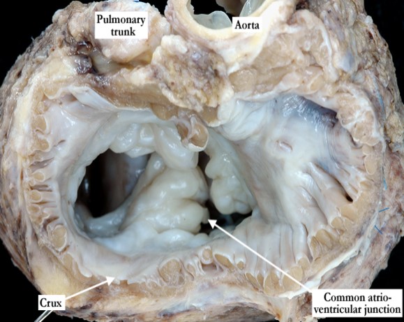
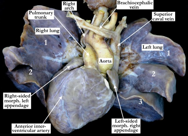
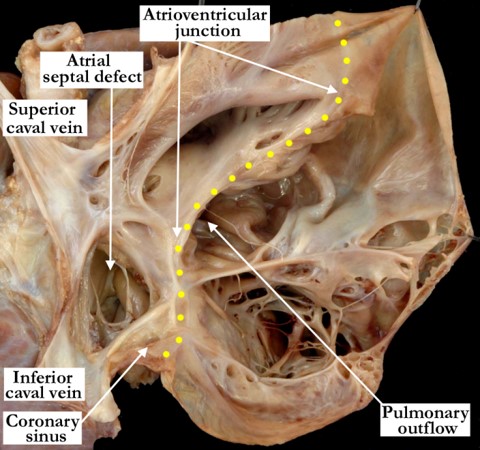
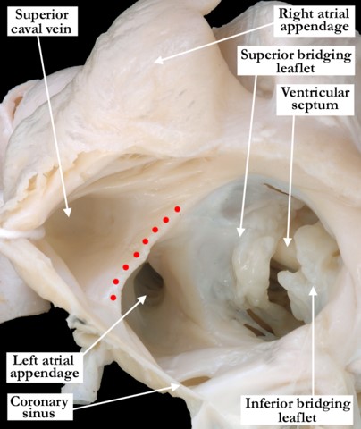
This view of the base of the heart demonstrates bilateral morphologically right atrial appendages. The pectinate muscles encircle the common atrioventricular junction on both sides, meeting at the crux. Note that a tongue of leaflet tissue joins together the bridging leaflets of the common valve, producing separate valvar orifice for the right and left ventricles.

[/restab][restab title=”Echocardiogram”]Content for Echocardiogram[/restab][restab title=”Computerized Axial Tomography”]Content for Computerized Axial Tomography[/restab][restab title=”MRI”]MRI[/restab][restab title=”Angiography”]Content for Angiography[/restab][restab title=”Intraoperative Videos”]Content for Intraoperative Videos[/restab][restab title=”Other”]Other[/restab][/restabs]

01.05.03 – Double outlet left ventricle

IPCCC Term DOUBLE OUTLET LEFT VENTRICLE

IPCCC Code 01.05.03

ICD-11 Code PENDING

Synonyms PENDING

Abbreviations PENDING

IPCCC Definition A congenital cardiovascular malformation in which both great arteries arise entirely or predominantly from the morphologically left ventricle.

[restabs alignment=”osc-tabs-right” pills=”nav-pills” responsive=”true” icon=”true” text=”More”][restab title=”Anatomic Specimen” active=”active”]

01-05-03 The left ventricle has been opened in clam-shell fashion, and the heart is viewed posteriorly. The mitral valve guards the inlet of the left ventricle. There is double outlet (red arrows) from the left ventricle, with both the aortic and pulmonary valves supported by completely muscular subarterial infundibulums. The interventricular communication is subpulmonary and, when viewed from the right ventricle, has exclusively muscular borders.

[/restab][restab title=”Echocardiogram”]Content for Echocardiogram[/restab][restab title=”Computerized Axial Tomography”]Content for Computerized Axial Tomography[/restab][restab title=”MRI”]MRI[/restab][restab title=”Angiography”]Content for Angiography[/restab][restab title=”Intraoperative Videos”]Content for Intraoperative Videos[/restab][restab title=”Other”]Other[/restab][/restabs]

01.01.40 + 07.13.04 – Double outlet right ventricle with subaortic ventricular septal defect without pulmonary stenosis

IPCCC Term DOUBLE OUTLET RIGHT VENTRICLE WITH SUBAORTIC VENTRICULAR SEPTAL DEFECT WITHOUT PULMONARY STENOSIS

IPCCC Code 01.01.40 + 07.13.04

ICD-11 Code PENDING

Synonyms PENDING

Abbreviations PENDING

IPCCC Definition A congenital cardiovascular malformation that is a variant of double outlet right ventricle with concordant atrioventricular connections, a subaortic ventricular septal defect, and unobstructed pulmonary outflow tract.

[restabs alignment=”osc-tabs-right” pills=”nav-pills” responsive=”true” icon=”true” text=”More”][restab title=”Anatomic Specimen” active=”active”]

01-01-40 and 07-13-04 This heart is viewed anteriorly and has been sectioned in a long axis plane. Both arterial trunks take origin predominantly from the right ventricle in the setting of a subaortic interventricular communication and an unobstructed pulmonary outflow tract. The defect, which opens to the right ventricle between the limbs of the septal band, is restrictive and perimembranous, since there is fibrous continuity between the leaflets of the aortic and tricuspid valves. The aortic valve is also in fibrous continuity with the anterior leaflet of the mitral valve.

[/restab][restab title=”Echocardiogram”]Content for Echocardiogram[/restab][restab title=”Computerized Axial Tomography”]Content for Computerized Axial Tomography[/restab][restab title=”MRI”]MRI[/restab][restab title=”Angiography”]Content for Angiography[/restab][restab title=”Intraoperative Videos”]Content for Intraoperative Videos[/restab][restab title=”Other”]Other[/restab][/restabs]

01.01.17 + 07.13.02 – Double outlet right ventricle with doubly committed ventricular septal defect & pulmonary stenosis

IPCCC Term DOUBLE OUTLET RIGHT VENTRICLE WITH DOUBLY COMMITTED VENTRICULAR SEPTAL DEFECT & PULMONARY STENOSIS

IPCCC Code 01.01.17 + 07.13.02

ICD-11 Code PENDING

Synonyms PENDING

Abbreviations PENDING

IPCCC Definition A congenital cardiovascular malformation that is a variant of double outlet right ventricle with concordant atrioventricular connections, a doubly committed ventricular septal defect with absence or deficiency of the conal septum, and pulmonary outflow tract obstruction.

[restabs alignment=”osc-tabs-right” pills=”nav-pills” responsive=”true” icon=”true” text=”More”][restab title=”Anatomic Specimen” active=”active”]

01-01-17 and 07-13-02 The right ventricle is opened in clam-shell fashion and viewed anteriorly to show both arterial trunks arising from the ventricle, producing the ventriculo-arterial connection of double outlet right ventricle. In this heart, the outlet septum (black asterisk) is fibrous rather than muscular, due to the presence of fibrous continuity between the leaflets of the pulmonary and aortic valves (yellow double headed arrow). In this case, there is also pulmonary stenosis. The interventricular communication lies between the limbs of the septal band, with fibrous continuity between the tricuspid and aortic valves, rendering the defect perimembranous. The defect is doubly committed, opening beneath both arterial outlets.

[/restab][restab title=”Echocardiogram”]Content for Echocardiogram[/restab][restab title=”Computerized Axial Tomography”]Content for Computerized Axial Tomography[/restab][restab title=”MRI”]MRI[/restab][restab title=”Angiography”]Content for Angiography[/restab][restab title=”Intraoperative Videos”]Content for Intraoperative Videos[/restab][restab title=”Other”]Other[/restab][/restabs]

03.01.03 – Total mirror imagery (situs inversus totalis)

IPCCC Term TOTAL MIRROR IMAGERY (SITUS INVERSUS TOTALIS)

IPCCC Code 03.01.03

ICD-11 Code PENDING

Synonyms PENDING

Abbreviations PENDING

IPCCC Definition A congenital malformation in which there is complete mirror-imaged arrangement of the internal organs along the left-right axis of the body.

[restabs alignment=”osc-tabs-right” pills=”nav-pills” responsive=”true” icon=”true” text=”More”][restab title=”Anatomic Specimen” active=”active”]

03-01-03 This anterior view of the heart and lungs demonstrates complete mirror-imaged arrangement of the internal organs of the thorax along the left-right axis of the body. The bronchial morphology (not seen here) was mirror-imaged with a trilobed lung on the left and a bilobed lung on the right. The cardiac apex is to the right, the caval veins are to the left with a mirror-imaged brachiocephalic vein. The atriums and ventricles are mirror-imaged and there is mirror-imaged spiraling of the arterial trunks. The aortic arch is to the right with mirror-imaged branching of the brachiocephalic arteries.

[/restab][restab title=”Echocardiogram”]Content for Echocardiogram[/restab][restab title=”Computerized Axial Tomography”]Content for Computerized Axial Tomography[/restab][restab title=”MRI”]MRI[/restab][restab title=”Angiography”]Content for Angiography[/restab][restab title=”Intraoperative Videos”]Content for Intraoperative Videos[/restab][restab title=”Other”]Other[/restab][/restabs]

02.03.03 – Crisscross heart (twisted atrioventricular connections)

IPCCC Term CRISSCROSS HEART (TWISTED ATRIOVENTRICULAR CONNECTIONS)

IPCCC Code 02.03.03

ICD-11 Code PENDING

Synonyms PENDING

Abbreviations PENDING

IPCCC Definition A congenital cardiac malformation in which the vectors of the atrioventricular inflow tracts are approximately orthogonal (perpendicular) to each other.

[restabs alignment=”osc-tabs-right” pills=”nav-pills” responsive=”true” icon=”true” text=”More”][restab title=”Anatomic Specimen” active=”active”]

02-03-03 This heart is viewed in anatomic position and is sectioned in a long axis plane demonstrating the features of a crisscross heart (twisted atrioventricular connections). In this heart, the vectors of the atrioventricular inflow tracts are approximately orthogonal or perpendicular to each other. The right atrioventricular connection is marked with the red arrow and the left with the yellow arrow. This heart has concordant atrioventricular connections and discordant ventriculo-arterial connections.

[/restab][restab title=”Echocardiogram”]Content for Echocardiogram[/restab][restab title=”Computerized Axial Tomography”]Content for Computerized Axial Tomography[/restab][restab title=”MRI”]MRI[/restab][restab title=”Angiography”]Content for Angiography[/restab][restab title=”Intraoperative Videos”]Content for Intraoperative Videos[/restab][restab title=”Other”]Other[/restab][/restabs]

01.01.56 – Tetralogy of Fallot with absent pulmonary valve

IPCCC Term TETRALOGY OF FALLOT WITH ABSENT PULMONARY VALVE

IPCCC Code 01.01.56

ICD-11 Code PENDING

Synonyms PENDING

Abbreviations PENDING

IPCCC Definition A congenital cardiovascular malformation that is a variant of tetralogy of Fallot in which the ventriculo-arterial junction of the right ventricle with the pulmonary trunk features an atypical valve with rudimentary leaflets (cusps) which do not coapt.

[restabs alignment=”osc-tabs-right” pills=”nav-pills” responsive=”true” icon=”true” text=”More”][restab title=”Anatomic Specimen” active=”active”]

[su_carousel source=”media: 6261,6262″ limit=”27″ width=”460″ height=”510″ responsive=”no” items=”1″ title=”no” centered=”no” mousewheel=”no” autoplay=”0″] This heart demonstrates tetralogy of Fallot with absent pulmonary valve. In Image #1, the right ventricle has been opened in clam-shell fashion showing the right ventricular inlet and outlet components with the aortic valve overriding the perimembranous ventricular septal defect. Note the anterior malalignment of the outlet septum. In Image #2, the right ventricular outflow tract is viewed from the anterior aspect with an elongated outlet component secondary to the anterior deviation of the outlet septum. The pulmonary valve consists of rudimentary, nodular, valve tissue that fails to coapt. This so-called absent pulmonary valve produces varying degrees of both pulmonary stenosis and regurgitation, often with physiologic dilation of the pulmonary trunk and right and left pulmonary arteries. The arterial duct (not seen) in this heart is absent, which is often the case. The aortic arch extends to the right with mirror image branching of the brachiocephalic arteries. (yellow star – trachea)

[/restab][restab title=”Echocardiogram”]Content for Echocardiogram.[/restab][restab title=”Computerized Axial Tomography”]Content for Computerized Axial Tomography.[/restab][restab title=”Angiography”]Content for Angiography.[/restab][restab title=”Intraoperative Videos”]Content for Intraoperative Videos.[/restab][/restabs]

07.01.06 – Parchment right ventricle (including Uhl anomaly)

IPCCC Term PARCHMENT RIGHT VENTRICLE (INCLUDING UHL ANOMALY)

IPCCC Code 07.01.06

ICD-11 Code PENDING

Synonyms PENDING

Abbreviations PENDING

IPCCC Definition A congenital cardiac malformation in which there is almost complete absence of right ventricular myocardium and preserved septal and left ventricular myocardium.

[restabs alignment=”osc-tabs-right” pills=”nav-pills” responsive=”true” icon=”true” text=”More”][restab title=”Anatomic Specimen” active=”active”]

07-01-06 The right atrium and ventricle have been opened in clam-shell fashion and are viewed from the antero-lateral aspect. There is almost complete absence of the right ventricular myocardium in this heart with an unguarded right ventricular inlet (yellow dots mark the atrioventricular junction) and pulmonary atresia with intact ventricular septum. The septal and left ventricular myocardium are preserved (left ventricle not shown).

[/restab][restab title=”Echocardiogram”]Content for Echocardiogram[/restab][restab title=”Computerized Axial Tomography”]Content for Computerized Axial Tomography[/restab][restab title=”MRI”]MRI[/restab][restab title=”Angiography”]Content for Angiography[/restab][restab title=”Intraoperative Videos”]Content for Intraoperative Videos[/restab][restab title=”Other”]Other[/restab][/restabs]

07.14.02 – Gerbode defect

IPCCC Term GERBODE DEFECT

IPCCC Code 07.14.02

ICD-11 Code PENDING

Synonyms PENDING

Abbreviations PENDING

IPCCC Definition A deficiency of the atrioventricular component of the membranous septum permitting shunting of blood from the left ventricle to the right atrium.

[restabs alignment=”osc-tabs-right” pills=”nav-pills” responsive=”true” icon=”true” text=”More”][restab title=”Anatomic Specimen” active=”active”]

07-14-02 This surgeon’s view of the right atrium and right atrioventricular junction shows a Gerbode defect. This defect is a deficiency of the atrioventricular component of the membranous septum that permits shunting of blood from the left ventricle to the right atrium.

[/restab][restab title=”Echocardiogram”]Content for Echocardiogram[/restab][restab title=”Computerized Axial Tomography”]Content for Computerized Axial Tomography[/restab][restab title=”MRI”]MRI[/restab][restab title=”Angiography”]Content for Angiography[/restab][restab title=”Intraoperative Videos”]Content for Intraoperative Videos[/restab][restab title=”Other”]Other[/restab][/restabs]

05.06.03 – Common atrium with common atrioventricular junction

IPCCC Term COMMON ATRIUM WITH COMMON ATRIOVENTRICULAR JUNCTION

IPCCC Code 05.06.03

ICD-11 Code PENDING

Synonyms PENDING

Abbreviations PENDING

IPCCC Definition A congenital cardiac malformation in which there is complete absence of the interatrial septum in the setting of a common atrioventricular junction (common atrioventricular canal).

[restabs alignment=”osc-tabs-right” pills=”nav-pills” responsive=”true” icon=”true” text=”More”][restab title=”Anatomic Specimen” active=”active”]

05-06-03 The inferior, lateral wall of the right atrial appendage has been cut and folded away demonstrating a common atrioventricular junction that is guarded by a common atrioventricular valve. The superior and inferior bridging leaflets are seen crossing the interventricular septum and the coronary sinus is easily visualized within the inferior right atrium. The entire atrial septum is absent with the red dots marking the roof of the common atrial chamber. There are concordant atrioventricular connections with a morphologically right atrial appendage anteriorly and the narrow opening of the morphologically left atrial appendage viewed through the common atrium along its posterior aspect.

[/restab][restab title=”Echocardiogram”]Content for Echocardiogram[/restab][restab title=”Computerized Axial Tomography”]Content for Computerized Axial Tomography[/restab][restab title=”MRI”]MRI[/restab][restab title=”Angiography”]Content for Angiography[/restab][restab title=”Intraoperative Videos”]Content for Intraoperative Videos[/restab][restab title=”Other”]Other[/restab][/restabs]