01.03.03 Isomerism of left atrial appendages

IPCCC Term ISOMERISM OF LEFT ATRIAL APPENDAGES

IPCCC Code 01.03.03

ICD-11 Code PENDING

Synonyms PENDING

Abbreviations PENDING

IPCCC Definition A congenital cardiac malformation in which both atrial appendages have the morphology of a left atrial appendage.

[restabs alignment=”osc-tabs-right” pills=”nav-pills” responsive=”true” icon=”true” text=”More”][restab title=”Anatomic Specimen” active=”active”]

01-03-03 
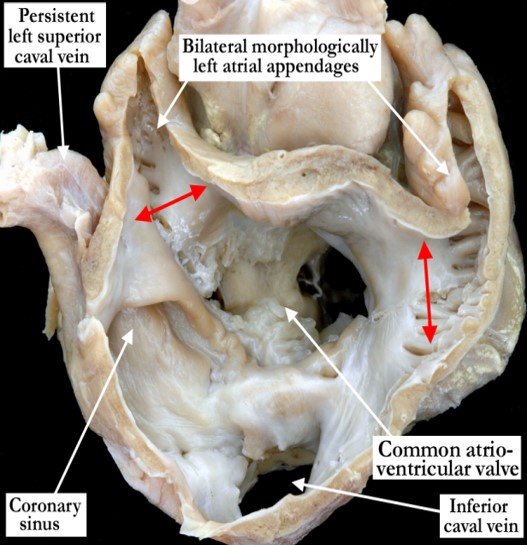
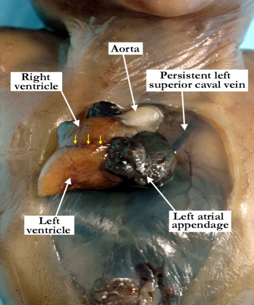
This view of the base of the heart demonstrates bilateral morphologically left atrial appendages. The pectinate muscles are confined within the tubular appendages bilaterally and both have a narrow junction (double headed red arrows) with the body of the atriums. The vestibules are smooth circumferentially to the crux on both sides. Note the common atrioventricular junction and the persistent left superior caval vein draining to the coronary sinus.

[/restab][restab title=”Echocardiogram”]Content for Echocardiogram[/restab][restab title=”Computerized Axial Tomography”]Content for Computerized Axial Tomography[/restab][restab title=”MRI”]MRI[/restab][restab title=”Angiography”]Content for Angiography[/restab][restab title=”Intraoperative Videos”]Content for Intraoperative Videos[/restab][restab title=”Other”]Other[/restab][/restabs]

02.01.01 Extrathoracic Heart

IPCCC Term EXTRATHORACIC HEART

IPCCC Code 02.01.01

ICD-11 Code PENDING

Synonyms PENDING

Abbreviations PENDING

IPCCC Definition A congenital cardiovascular malformation in which the heart is at least partially outside of the thorax.

[restabs alignment=”osc-tabs-right” pills=”nav-pills” responsive=”true” icon=”true” text=”More”][restab title=”Anatomic Specimen” active=”active”]

02-01-01 This is an anterior view of the chest wall in a stillborn fetus at 18 weeks gestation with exteriorization of the heart. The heart is exteriorized because of the deficiency of the surrounding thoraco-abdominal wall. The cardiac apex is to the right, the left atrium is markedly dilated and the persistent left superior caval vein is easily visualized through the thin thoracic membrane. Internal examination revealed tetralogy of Fallot ,with a small right ventricle that did not extend to the apex of the heart. Note the anterior interventricular coronary artery marked with yellow arrows.

[/restab][restab title=”Echocardiogram”]Content for Echocardiogram[/restab][restab title=”Computerized Axial Tomography”]Content for Computerized Axial Tomography[/restab][restab title=”MRI”]MRI[/restab][restab title=”Angiography”]Content for Angiography[/restab][restab title=”Intraoperative Videos”]Content for Intraoperative Videos[/restab][restab title=”Other”]Other[/restab][/restabs]

06.02.03 Dysplasia of the mitral valve

IPCCC Term DYSPLASIA OF THE MITRAL VALVE

IPCCC Code 06.02.03

ICD-11 Code PENDING

Synonyms PENDING

Abbreviations PENDING

IPCCC Definition A congenital cardiac malformation that includes any structural abnormality of the mitral valvar leaflet(s), commonly consisting of leaflet thickening and restricted mobility.

[restabs alignment=”osc-tabs-right” pills=”nav-pills” responsive=”true” icon=”true” text=”More”][restab title=”Anatomic Specimen” active=”active”]

[su_carousel source=”media: 5377,5378″ limit=”27″ width=”460″ height=”510″ responsive=”no” items=”1″ title=”no” centered=”no” mousewheel=”no” autoplay=”0″] In Image #1, this postero-superior view of the left atrioventricular junction demonstrates a stenotic, thickened, dysplastic mitral valve. The inferior horn of the flap valve is marked with red dots. In Image #2, the left atrioventricular junction is opened and viewed posteriorly, demonstrating a thickened, dysplastic mitral valve. All of the leaflets are supported by thickened, fused tendinous cords.

[/restab][restab title=”Echocardiogram”]Content for Echocardiogram.[/restab][restab title=”Computerized Axial Tomography”]Content for Computerized Axial Tomography.[/restab][restab title=”Angiography”]Content for Angiography.[/restab][restab title=”Intraoperative Videos”]Content for Intraoperative Videos.[/restab][/restabs]

06.01.25 Congenital tricuspid regurgitation

IPCCC Term CONGENITAL TRICUSPID REGURGITATION

IPCCC Code 06.01.25

ICD-11 Code PENDING

Synonyms PENDING

Abbreviations PENDING

IPCCC Definition A congenital cardiac finding in which there is backward flow through the tricuspid valve.

[restabs alignment=”osc-tabs-right” pills=”nav-pills” responsive=”true” icon=”true” text=”More”][restab title=”Anatomic Specimen” active=”active”]

[su_carousel source=”media: 5375,5376″ limit=”27″ width=”460″ height=”510″ responsive=”no” items=”1″ title=”no” centered=”no” mousewheel=”no” autoplay=”0″] In Image #1, the right atrium and ventricle are opened in clam-shell fashion and viewed anteriorly in anatomic position. The right atrioventricular junction is guarded by a dysplstic and regurgitant tricuspid valve. All of the leaflets are redundant with a dilated, ballooned appearance in some areas. The tendinous cords are elongated. In image #2, the superior lateral view of the tricuspid valve inlet demonstrates a balloon-like anterior leaflet. The tricuspid valve does not occlude properly the atrioventricular orifice.

[/restab][restab title=”Echocardiogram”]Content for Echocardiogram.[/restab][restab title=”Computerized Axial Tomography”]Content for Computerized Axial Tomography.[/restab][restab title=”Angiography”]Content for Angiography.[/restab][restab title=”Intraoperative Videos”]Content for Intraoperative Videos.[/restab][/restabs]

06.01.05 Overriding tricuspid valve

IPCCC Term OVERRIDING TRICUSPID VALVE

IPCCC Code 06.01.05

ICD-11 Code PENDING

Synonyms PENDING

Abbreviations PENDING

IPCCC Definition A congenital cardiac malformation in which the tricuspid valve annulus lies in part above both the right and left ventricles.

[restabs alignment=”osc-tabs-right” pills=”nav-pills” responsive=”true” icon=”true” text=”More”][restab title=”Anatomic Specimen” active=”active”]

[su_carousel source=”media: 5373,5374″ limit=”27″ width=”460″ height=”510″ responsive=”no” items=”1″ title=”no” centered=”no” mousewheel=”no” autoplay=”0″] Image #1 the right atrium and ventricle are opened in clam-shell fashion and are viewed anteriorly. The right atrioventricular junction is shown with a perimembranous ventricular septal defect beneath the septal leaflet of the tricuspid valve (red dots). The defect essentially divides the septal leaflet with the portion adjacent to the hinge point of the valve overriding the ventricular septum. Image #2 is a posterior view of the left ventriculo-arterial junction and demonstrates the overriding tricuspid valve with a portion of the tricuspid valve annulus lying in part above the left ventricle. Note that there are no tendinous cords supporting the tricuspid valve within the left ventricle.

[/restab][restab title=”Echocardiogram”]Content for Echocardiogram.[/restab][restab title=”Computerized Axial Tomography”]Content for Computerized Axial Tomography.[/restab][restab title=”Angiography”]Content for Angiography.[/restab][restab title=”Intraoperative Videos”]Content for Intraoperative Videos.[/restab][/restabs]

05.05.03 Interatrial communication through coronary sinus orifice

IPCCC Term INTERATRIAL COMMUNICATION THROUGH CORONARY SINUS ORIFICE

IPCCC Code 05.05.03

ICD-11 Code PENDING

Synonyms PENDING

Abbreviations PENDING

IPCCC Definition A congenital cardiovascular malformation in which there is a communication between the left atrium and the coronary sinus allowing interatrial communication through the coronary sinus ostium.

[restabs alignment=”osc-tabs-right” pills=”nav-pills” responsive=”true” icon=”true” text=”More”][restab title=”Anatomic Specimen” active=”active”]

[su_carousel source=”media: 5371,5372″ limit=”27″ width=”460″ height=”510″ responsive=”no” items=”1″ title=”no” centered=”no” mousewheel=”no” autoplay=”0″] Image #1 is viewed anteriorly demonstrating the septal surfaces of an opened right atrium and ventricle with a large coronary sinus orifice, acting as an interatrial communication. The oval foramen was restrictive with premature closure. Image #2 is a posterior view of the left side of the same heart. The left atrium and ventricle are hypoplastic and the pulmonary veins are normally connected. The typical horseshoe attachment of the flap valve within the left atrium is marked with red dots. There is a large interatrial communication at the coronary sinus. In this case the interatrial communication is not associated with a persistent left superior caval vein.

[/restab][restab title=”Echocardiogram”]Content for Echocardiogram.[/restab][restab title=”Computerized Axial Tomography”]Content for Computerized Axial Tomography.[/restab][restab title=”Angiography”]Content for Angiography.[/restab][restab title=”Intraoperative Videos”]Content for Intraoperative Videos.[/restab][/restabs]

05.02.02 Supravalvar or intravalvar mitral ring

IPCCC Term Supravalvar or intravalvar mitral ring

IPCCC Code 05.02.02

ICD-11 Code PENDING

Synonyms PENDING

Abbreviations PENDING

IPCCC Definition A congenital cardiac malformation in which a ridge of tissue is attached or integral to the atrial side of the mitral valvar leaflet(s).

[restabs alignment=”osc-tabs-right” pills=”nav-pills” responsive=”true” icon=”true” text=”More”][restab title=”Anatomic Specimen” active=”active”]

[su_carousel source=”media: 5369,5370″ limit=”27″ width=”850″ height=”620″ responsive=”no” items=”1″ title=”no” centered=”no” mousewheel=”no”] Left Panel: This postero-superior view of the left atrioventricular junction shows a prominent, supravalvar ring (yellow dots) associated with the atrial aspect of the mitral valve. This ring is above the annulus or hinge point of the valve. Right Panel: The left atrioventricular junction is opened and viewed posteriorly, demonstrating a thickened mitral valve with thickened fused tendinous cords. Within the funnel of the mitral valve there is a prominent intravalvar ring (yellow dots).

[/restab][restab title=”Echocardiogram”]Content for Echocardiogram.[/restab][restab title=”Computerized Axial Tomography”]Content for Computerized Axial Tomography.[/restab][restab title=”Angiography”]Content for Angiography.[/restab][restab title=”Intraoperative Videos”]Content for Intraoperative Videos.[/restab][/restabs]

05.02.01 Divided left atrium (cor triatriatum sinister)

IPCCC Term Divided left atrium (cor triatriatum sinister)

IPCCC Code 05.02.01

ICD-11 Code PENDING

Synonyms PENDING

Abbreviations PENDING

IPCCC Definition A congenital cardiac malformation in which there is a partition that divides the left atrium into a posterior chamber that receives some or all of the pulmonary veins and an anterior chamber that communicates with the left atrial appendage and atrioventricular junction (usually the mitral valve).

[restabs alignment=”osc-tabs-right” pills=”nav-pills” responsive=”true” icon=”true” text=”More”][restab title=”Anatomic Specimen” active=”active”]

[su_carousel source=”media: 5367,5368″ limit=”27″ width=”720″ height=”540″ responsive=”no” items=”1″ title=”no” centered=”no” mousewheel=”no”] The left panel shows a posterior view of an opened left atrial chamber and the pulmonary veins connecting to a posterior chamber. The yellow dashed arrow illustrates the path of the pulmonary venous drainage from the posterior chamber to the left atrial vestibule. The opened mitral valve guards the left atrioventricular junction. In the right panel this postero-superior view shows the partition dividing the posterior left atrial chamber from the atrial vestibule. The pulmonary veins drain into the atrial vestibule via a small opening (yellow dots) in the floor of the partition that divides the left atrium.

[/restab][restab title=”Echocardiogram”]Content for Echocardiogram.[/restab][restab title=”Computerized Axial Tomography”]Content for Computerized Axial Tomography.[/restab][restab title=”Angiography”]Content for Angiography.[/restab][restab title=”Intraoperative Videos”]Content for Intraoperative Videos.[/restab][/restabs]

09.16.18 Congenital supravalvar aortic stenosis

IPCCC Term Congenital supravalvar aortic stenosis

IPCCC Code 09.16.18

ICD-11 Code PENDING

Synonyms PENDING

Abbreviations PENDING

IPCCC Definition A congenital cardiovascular malformation with narrowing of the aorta at the level of the sinotubular junction which may extend into the ascending aorta.

[restabs alignment=”osc-tabs-right” pills=”nav-pills” responsive=”true” icon=”true” text=”More”][restab title=”Anatomic Specimen” active=”active”]

[su_carousel source=”media: 4986,4987″ limit=”27″ width=”460″ height=”510″ responsive=”no” items=”1″ title=”no” centered=”no” mousewheel=”no”] Image # 1 is an anterior superior view of the aortic root as it exits the ventricular mass there is marked dilation of the sinuses of Valsalva secondary to a fibrous membrane present internally at the level of the sinotubular junction (red dots).In image # 2 the left ventricular outflow tract and the aortic root shows subvalvar and supravalvar aortic stenosis in this patient noted to have Shone’s syndrome. The subvalvar stenosis is caused by a prominent fibrous ridge that is circumferential and extends over the aorta to mitral fibrous continuity. At the sinotubular junction (red dots) there is a fibrous shelf accounting for the distal valvar stenosis. Although this is the lesion that is commonly defined as being “supravalvar”, it is in fact a valvar lesion since the “commissures”, or the peripheral attachments of the leaflets, are part of the valvar mechanism. The sinuses of Valsalva are markedly dilated (not seen in this image) with the right and non-adjacent leaflets tethered to the fibrous shelf.

[/restab][restab title=”Echocardiogram”]Content for Echocardiogram.[/restab][restab title=”Computerized Axial Tomography”]Content for Computerized Axial Tomography.[/restab][restab title=”Angiography”]Content for Angiography.[/restab][restab title=”Intraoperative Videos”]Content for Intraoperative Videos.[/restab][/restabs]