05.01.12 – Congenital giant right atrium

IPCCC Term CONGENITAL GIANT RIGHT ATRIUM

IPCCC Code 05.01.12

ICD-11 Code PENDING

Synonyms PENDING

Abbreviations PENDING

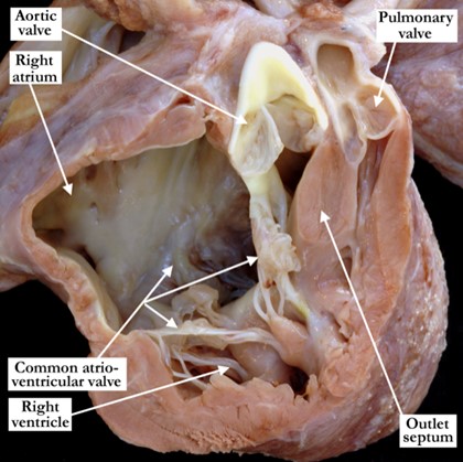
IPCCC Definition A congenital cardiac malformation in which the right atrium is severely dilated. This is an isolated finding not secondary to abnormalities of the tricuspid valve or right ventricle.[restabs alignment=”osc-tabs-right” pills=”nav-pills” responsive=”true” icon=”true” text=”More”][restab title=”Anatomic Specimen” active=”active”]

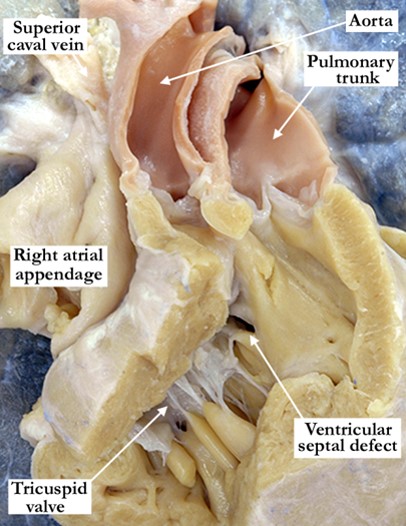
[su_carousel source=”media: 4982,4983″ limit=”27″ width=”460″ height=”510″ responsive=”no” items=”1″ title=”no” centered=”no” mousewheel=”no” autoplay=”0″]Image # 1 demonstrates he anterior anatomic view of a grossly dilated right atrium. Image # 2 shows the heart an anterior view of the heart with an opened right atrium, demonstrating marked dilation of the appendage and atrial chamber. The oval foramen is probe patent without appreciable dilation of the flap valve. There are concordant atrioventricular and ventriculo-arterial connections without abnormalities of the atrioventricular or arterial valves.
[/restab][restab title=”Echocardiogram”]Content for Echocardiogram.[/restab][restab title=”Computerized Axial Tomography”]Content for Computerized Axial Tomography.[/restab][restab title=”Angiography”]Content for Angiography.[/restab][restab title=”Intraoperative Videos”]Content for Intraoperative Videos.[/restab][/restabs]

01.01.20 – Atrioventricular septal defect and tetralogy of Fallot (atrioventricular canal and tetralogy of Fallot)

IPCCC Term ATRIOVENTRICULAR SEPTAL DEFECT AND TETRALOGY OF FALLOT (ATRIOVENTRICULAR CANAL AND TETRALOGY OF FALLOT)

IPCCC Code 01.01.20

ICD-11 Code PENDING

Synonyms PENDING

Abbreviations PENDING

IPCCC Definition A congenital cardiac malformation with both an atrioventricular septal defect (atrioventricular canal defect) and tetralogy of Fallot.[restabs alignment=”osc-tabs-right” pills=”nav-pills” responsive=”true” icon=”true” text=”More”][restab title=”Anatomic Specimen” active=”active”]

01-01-20-atrioventricular-septal-defect-and-tetralogy-of-fallot-atrioventricular-canal-and-tetralogy-of-fallotThis heart is viewed in an anterior anatomic position and has been sectioned to illustrate the left and right ventricular outflow tracts and common atrioventricular valve. The specimen is photographed from the front, simulating the subcostal long-axis echocardiographic plane. There is antero-cephalad deviation of the outlet septum producing severe subpulmonary infundibular and valvar stenosis. The aortic valve overrides the interventricular septum in this heart with tetralogy of Fallot. There is also an atrioventricular septal defect with a common atrioventricular junction and valve.
[/restab][restab title=”Echocardiogram”]Content for Echocardiogram.[/restab][restab title=”Computerized Axial Tomography”]Content for Computerized Axial Tomography.[/restab][restab title=”Angiography”]Content for Angiography.[/restab][restab title=”Intraoperative Videos”]Content for Intraoperative Videos.[/restab][/restabs]

06.07.05 – Atrioventricular septal defect with ventricular imbalance: dominant right ventricle, hypoplastic left ventricle

IPCCC Term ATRIOVENTRICULAR SEPTAL DEFECT WITH VENTRICULAR IMBALANCE: DOMINANT RIGHT VENTRICLE, HYPOPLASTIC LEFT VENTRICLE

IPCCC Code 06.07.05

ICD-11 Code PENDING

Synonyms PENDING

Abbreviations PENDING

IPCCC Definition A congenital cardiac malformation that is a form of atrioventricular septal defect (atrioventricular canal defect) with the right ventricle significantly larger than the left.

[restabs alignment=”osc-tabs-right” pills=”nav-pills” responsive=”true” icon=”true” text=”More”][restab title=”Anatomic Specimen” active=”active”]

06-07-05-atrioventricular-septal-defect-with-ventricular-imbalance This heart has been sectioned to demonstrate the short axis of the ventricular mass. It is photographed to show the view from the apex looking towards the base. There is an atrioventricular septal defect with a common atrioventricular junction. The common atrioventricular valve is committed predominately to the right ventricle, producing right ventricular dominance. The left ventricle is hypoplastic, with excessive trabeculations and closely adjacent papillary muscles. The superior and inferior portions of the deficient muscular ventricular septum are marked with the black stars.

[/restab][restab title=”Echocardiogram”]Content for Echocardiogram.[/restab][restab title=”Computerized Axial Tomography”]Content for Computerized Axial Tomography.[/restab][restab title=”Angiography”]Content for Angiography.[/restab][restab title=”Intraoperative Videos”]Content for Intraoperative Videos.[/restab][/restabs]

06.07.06 – Atrioventricular septal defect with ventricular imbalance: dominant left ventricle, hypoplastic right ventricle

IPCCC Term ATRIOVENTRICULAR SEPTAL DEFECT WITH VENTRICULAR IMBALANCE: DOMINANT LEFT VENTRICLE, HYPOPLASTIC RIGHT VENTRICLE

IPCCC Code 06.07.06

ICD-11 Code PENDING

Synonyms PENDING

Abbreviations PENDING

IPCCC Definition A congenital cardiac malformation that is a variant of an atrioventricular septal defect (atrioventricular canal defect) with the left ventricle significantly larger than the right.

[restabs alignment=”osc-tabs-right” pills=”nav-pills” responsive=”true” icon=”true” text=”More”][restab title=”Anatomic Specimen” active=”active”]

06-07-06-atrioventricular-septal-defect This heart has been sectioned to demonstrate the short axis of the ventricular mass. It is photographed to show the view from the apex looking towards the base. There is an atrioventricular septal defect with a common atrioventricular junction. The common atrioventricular valve is committed predominately to the left ventricle, resulting on left ventricular dominance. The papillary muscles (black stars) are close together within the left ventricle, which shows excessive trabeculations. The zone of apposition between the superior and inferior bridging leaflets is marked with the yellow lines. The right ventricular cavity is hypoplastic as is the right component of the common atrioventricular valve.

[/restab][restab title=”Echocardiogram”]Content for Echocardiogram.[/restab][restab title=”Computerized Axial Tomography”]Content for Computerized Axial Tomography.[/restab][restab title=”Angiography”]Content for Angiography.[/restab][restab title=”Intraoperative Videos”]Content for Intraoperative Videos.[/restab][/restabs]

02.06.05 – Aortic root directly leftward to pulmonary root (side-by-side)

IPCCC Term AORTIC ROOT DIRECTLY LEFTWARD TO PULMONARY ROOT (SIDE-BY-SIDE)

IPCCC Code 02.06.05

ICD-11 Code PENDING

Synonyms PENDING

Abbreviations PENDING

IPCCC Definition A congenital cardiovascular malformation in which the aortic root or its remnant is positioned directly to the left of the pulmonary root or its remnant.

[restabs alignment=”osc-tabs-right” pills=”nav-pills” responsive=”true” icon=”true” text=”More”][restab title=”Anatomic Specimen” active=”active”]

02-06-05-aortic-root-directly-leftward-to-pulmonary-root-side-by-side This anterior view demonstrates the heart in anatomic position. The cardiac apex, not shown in this image, is directed to the right. The aortic root is positioned side by side and directly leftward of the pulmonary root.

[/restab][restab title=”Echocardiogram”]Content for Echocardiogram.[/restab][restab title=”Computerized Axial Tomography”]Content for Computerized Axial Tomography.[/restab][restab title=”Angiography”]Content for Angiography.[/restab][restab title=”Intraoperative Videos”]Content for Intraoperative Videos.[/restab][/restabs]

02.06.01 – Aortic root directly rightward to pulmonary root (side-by-side)

IPCCC Term AORTIC ROOT DIRECTLY RIGHTWARD TO PULMONARY ROOT (SIDE-BY-SIDE)

IPCCC Code 02.06.01

ICD-11 Code PENDING

Synonyms PENDING

Abbreviations PENDING

IPCCC Definition A congenital cardiovascular malformation in which the aortic root or its remnant is positioned directly to the right of the pulmonary root or its remnant.

[restabs alignment=”osc-tabs-right” pills=”nav-pills” responsive=”true” icon=”true” text=”More”][restab title=”Anatomic Specimen” active=”active”]

02-06-01-aortic-root-directly-rightward A portion of the anterior, right ventricular free wall has been removed to demonstrate the relationships of the arterial trunks. The aortic root is positioned directly to the right of the pulmonary trunk in this heart with ventriculo-arterial connection of double outlet right ventricle. The arterial trunks are side by side and both are supported by a complete, subarterial muscular infundibulum. Note the right ventricular hypertrophy and the restrictive ventricular septal defect (interventricular communication).

[/restab][restab title=”Echocardiogram”]Content for Echocardiogram.[/restab][restab title=”Computerized Axial Tomography”]Content for Computerized Axial Tomography.[/restab][restab title=”Angiography”]Content for Angiography.[/restab][restab title=”Intraoperative Videos”]Content for Intraoperative Videos.[/restab][/restabs]

01.01.16 – Partial anomalous pulmonary venous connection of Scimitar type

IPCCC Term PARTIAL ANOMALOUS PULMONARY VENOUS CONNECTION OF SCIMITAR TYPE

IPCCC Code 01.01.16

ICD-11 Code PENDING

Synonyms PENDING

Abbreviations PENDING

IPCCC Definition A congenital cardiovascular malformation with partial anomalous pulmonary venous connection in which some of the pulmonary veins (usually the right pulmonary veins) connect anomalously to the inferior caval vein (inferior vena cava) or to the right atrium at the insertion of the inferior vena cava.

[restabs alignment=”osc-tabs-right” pills=”nav-pills” responsive=”true” icon=”true” text=”More”][restab title=”Anatomic Specimen” active=”active”]

[su_carousel source=”media: 4976,4977″ limit=”27″ width=”460″ height=”510″ responsive=”no” items=”1″ title=”no” centered=”no” mousewheel=”no”] This heart/lung block is obtained from a patient with Scimitar syndrome. Image # 1 shows the opened right atrium viewed from the front and slightly to the right. A probe has been placed in the solitary pulmonary vein draining the right lung, which opens anomalously into the inferior caval vein immediately above the diaphragm. Although not viewed in this image, the left pulmonary veins drained in the usual fashion to the left atrium.

[/restab][restab title=”Echocardiogram”]Content for Echocardiogram.[/restab][restab title=”Computerized Axial Tomography”]Content for Computerized Axial Tomography.[/restab][restab title=”Angiography”]Content for Angiography.[/restab][restab title=”Intraoperative Videos”]Content for Intraoperative Videos.[/restab][/restabs]]]>

02.01.04 – Mesocardia

IPCCC Term MESOCARDIA

IPCCC Code 02.01.04

ICD-11 Code PENDING

Synonyms PENDING

Abbreviations PENDING

IPCCC Definition A congenital cardiovascular malformation in which the heart is central or midline within the thorax.

[restabs alignment=”osc-tabs-right” pills=”nav-pills” responsive=”true” icon=”true” text=”More”][restab title=”Anatomic Specimen” active=”active”]

02-01-04-mesocardia The anterior portion of the sternum and ribs are removed to demonstrate the in situ position of the heart which lies in the midline with its apex pointing down. The yellow dots mark the limits of the diaphragmatic aspect of the pericardium.

[/restab][restab title=”Echocardiogram”]Content for Echocardiogram[/restab][restab title=”Computerized Axial Tomography”]Content for Computerized Axial Tomography[/restab][restab title=”MRI”]MRI[/restab][restab title=”Angiography”]Content for Angiography[/restab][restab title=”Intraoperative Videos”]Content for Intraoperative Videos[/restab][restab title=”Other”]Other[/restab][/restabs]