09.45.16 – Congenital coronary arterial fistula
IPCCC Term CONGENITAL CORONARY ARTERIAL FISTULA
IPCCC Code 09.45.16
ICD-11 Code PENDING
Synonyms PENDING
Abbreviations PENDING
IPCCC Definition A congenital cardiovascular malformation in which a coronary artery communicates, through an anomalous channel, with a cardiac chamber or with any segment of the pulmonary circulation.
[restabs alignment=”osc-tabs-right” pills=”nav-pills” responsive=”true” icon=”true” text=”More”][restab title=”Anatomic Specimen” active=”active”]
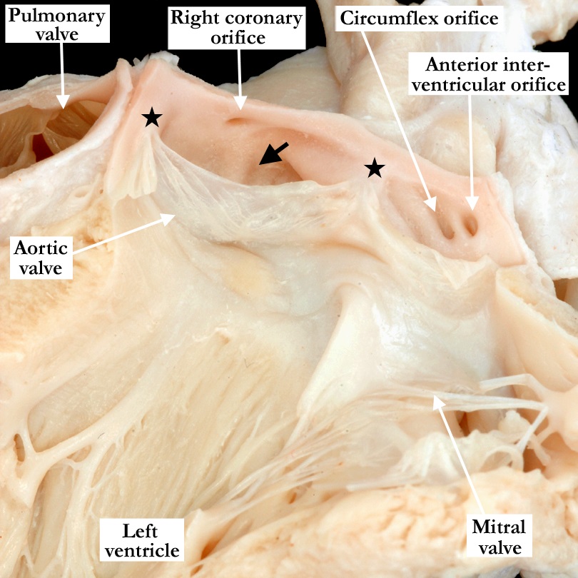
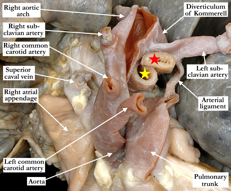
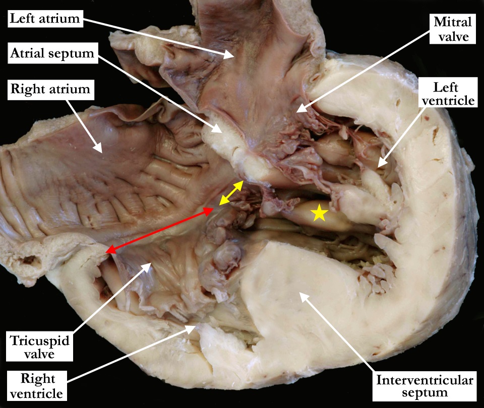
| [su_carousel source=”media: 6762,6763″ limit=”27″ width=”600″ height=”460″ responsive=”no” items=”1″ title=”no” centered=”no” mousewheel=”no” autoplay=”0″] | Image # 1: This anterior superior view of an explanted heart with hypoplastic left heart demonstrates multiple, ectatic coronary arteries over the epicardial surface with several communicating arteries between the right and left coronary circulations. These cross the anterior subpulmonary aspect of the right ventricle. The atretic origin of the left main coronary artery is marked with the black star with a long atretic segment extending to the large ectatic portion that bifurcates into the anterior interventricular and circumflex (black arrows) branches. There was aortic valve atresia (not shown). Image #2: A wedge of tissue is removed from the posterior aspect of the same heart seen in Image #1, demonstrating severe mitral valve stenosis and a hypoplastic left ventricle with a large coronary artery to left ventricular fistula. |
[/restab][restab title=”Echocardiogram”]Content for Echocardiogram.[/restab][restab title=”Computerized Axial Tomography”]Content for Computerized Axial Tomography.[/restab][restab title=”Angiography”]Content for Angiography.[/restab][restab title=”Intraoperative Videos”]Content for Intraoperative Videos.[/restab][/restabs]