|

(click image to
view original size) |
|
Derived Terms: |
|
|
AEPC: |
Normal heart (01.01.00) |
| |
Normal position-orientation of heart (02.01.00) |
| |
|
|
EACTS-STS: |
Normal heart (01.01.00) |
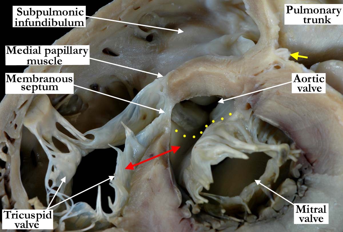
Modality: Anatomic specimen
Orientation: Basal short axis view (close
up)
Description: This heart has again been
dissected in the fashion of the previous short axis views (A010100-141a,
A010100-142a,
A010100-143a,
A010100-144a,
010100-145a and 010100-146a)
, and is shown in close-up fashion to demonstrate the relationship of the
tricuspid, mitral and aortic valves to the interventricular component of the
membranous septum. The membranous septum is located at the junction of the
inlet, outlet, and trabecular components of the muscular septum, with the
aorta deeply wedged between the mitral valve and the muscular septum
supporting the septal leaflet of the tricuspid valve. The membranous portion
of the interventricular septum establishes fibrous continuity between the
leaflets of the tricuspid and aortic valves. The area of aortic to mitral
fibrous continuity is demonstrated with yellow dots, and is seen to extend
to the border of the membranous septum. The subpulmonary muscular
infundibulum wraps around the aortic outflow from the left ventricle. The
mitral valve is lifted away from the septum, with the double headed red
arrow demonstrating how the right ventricular inlet is separated from the
subaortic outlet from the left ventricle. The yellow arrow indicates the
anterior interventricular coronary artery.
Contributor: Diane
E. Spicer, BS
Institution: The Congenital Heart
Institute of Florida (CHIF)
Image Label: A010100-147a
Source of Image: The Congenital Heart
Institute of Florida (CHIF)
Image Certification: pending
AWG Rating: pending
|
|