|
Derived Terms: |
|
|
AEPC: |
Normal heart (01.01.00) |
| |
Normal position-orientation of heart (02.01.00) |
| |
|
|
EACTS-STS: |
Normal heart (01.01.00) |
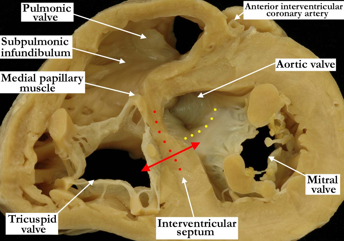
Modality: Anatomic specimen
Orientation: Basal short axis view
Description: This heart has been sectioned
in a similar manner to the previous short axis views (A010100-141a,
A010100-142a,
A010100-143a,
A010100-144a and
010100-145a) with the muscular interventricular septum (red dots)
further dissected toward the base of the heart. This view demonstrates
nicely how the right ventricular outlet wraps around the outlet of the left
ventricle, and shows well the deeply wedged position of the aortic valve
between the mitral valve and the septal components. The double-headed red
arrows show the portion of the interventricular septum that separates the
inlet component of the right ventricle from the outlet of the left
ventricle. The tricuspid valve is clearly septophilic, while the mitral
valve is septophobic, lacking any cordal attachments to the muscular
ventricular septum. The anterior, or aortic, leaflet of the mitral valve is
in fibrous continuity with the aortic valve (yellow dots).
Contributor: Diane
E. Spicer, BS
Institution: The Congenital Heart
Institute of Florida (CHIF)
Image Label: A010100-146a
Source of Image: The Congenital Heart
Institute of Florida (CHIF)
Image Certification: pending
AWG Rating: pending
|