|
||||||||
|
|
|||||||||
|
|||||||||
|
(click image to view original size) |
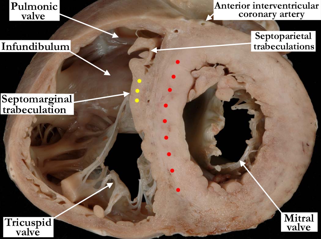
Modality: Anatomic specimen Orientation: Basal short axis view Description: Continuing the series of short axis sections shown in images A010100-141a, A010100-142a and A010100-143a, the cut shows how the septal surface of the left ventricle becomes smooth as it extends toward the aortic valve. The mitral valve continues to lift away from the septum. The septal surface of the right ventricle retains its coarse trabeculations, and the body of the septomarginal trabeculation (yellow dots) remains prominent. The right ventricle wraps around the left ventricular outlet component, this becoming more prominent as the images extend toward the base of the heart. Contributor: Diane E. Spicer, BS Institution: The Congenital Heart Institute of Florida (CHIF) Image Label: A010100-144a Source of Image: The Congenital Heart Institute of Florida (CHIF) Image Certification: pending AWG Rating: pending |
|||||||||||
AWG Page Certification: pending
|
Copyright ipccc-awg.net All Rights Reserved. Frontpage-Templates.org |