|
(click image to
view original size) |
|
Derived Terms: |
|
|
AEPC: |
Normal heart (01.01.00) |
| |
Normal position-orientation of heart (02.01.00) |
| |
|
|
EACTS-STS: |
Normal heart (01.01.00) |
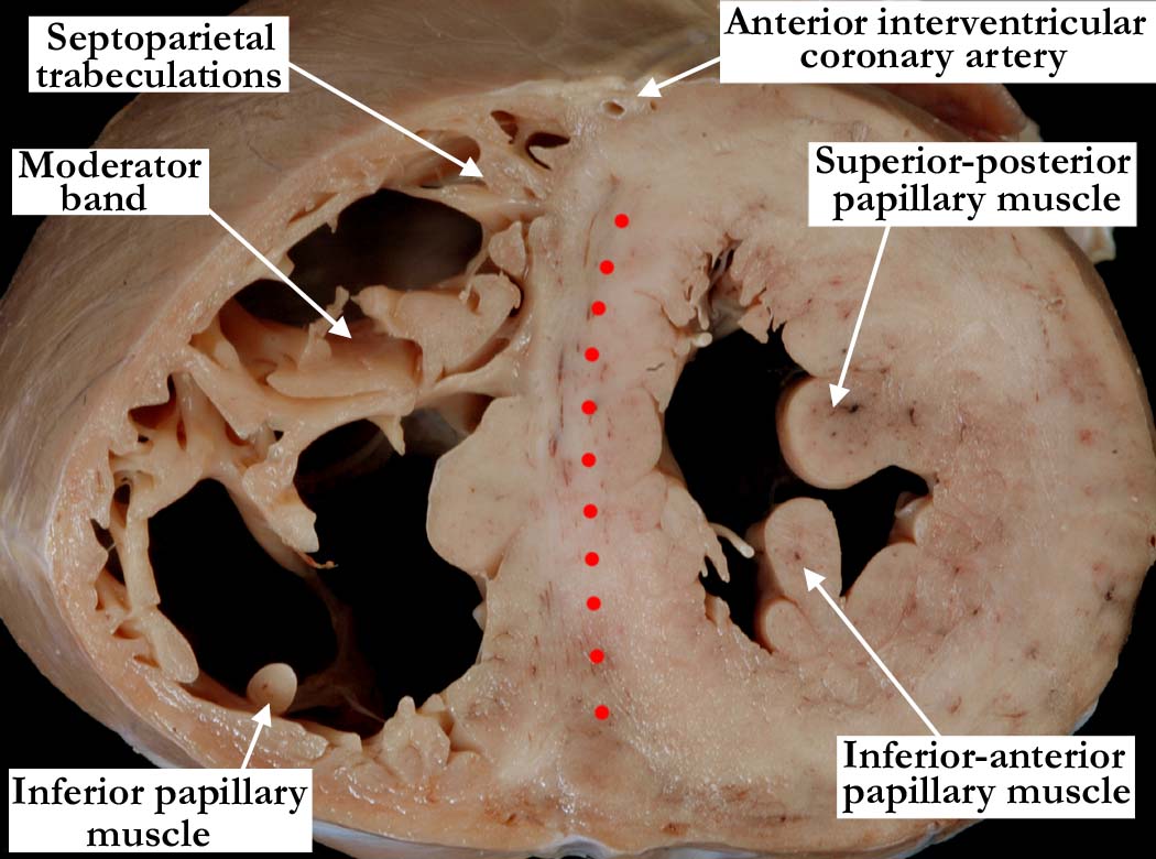
Modality: Anatomic specimen
Orientation: Apical short axis view
Description: This apical short axis view
is from the same heart as shown in image
A010100-141a, but is
taken closer to the base of the heart. It shows the coarsely trabeculated
septal surface, and the trabecular component extending between the septum
and the parietal wall of the right ventricle. On the inferior aspect of the
right ventricle is the inferior papillary muscle, which supports the zone of
apposition between the inferior, or mural, and the septal leaflets of the
tricuspid valve. The moderator band arises from a prominent right
ventricular trabeculation, the septomarginal trabeculation, and joins the
anterior papillary muscle, which supports the antero-superior leaflet of the
tricuspid valve. The section cuts through the junction of the inlet and
trabecular component of the left ventricle, showing the origins of the
superior-posterior and inferior-anterior papillary muscles of the mitral
valve, which arise from the free wall of the left ventricle. The muscular
interventricular septum is marked with red dots. It bulges to the right in
the normal heart.
Contributor: Diane
E. Spicer, BS
Institution: The Congenital Heart
Institute of Florida (CHIF)
Image Label: A010100-142a
Source of Image: The Congenital Heart
Institute of Florida (CHIF)
Image Certification: pending
AWG Rating: pending
|
|
