|
||||||||
|
|
|||||||||
|
|||||||||
|
(click image to view original size) |
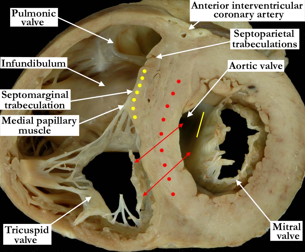
Modality: Anatomic specimen Orientation: Basal short axis view Description: The section continues the series shown in A010100-141a, A010100-142a, A010100-143a and A010100-144a. The right ventricular outlet, better described as the subpulmonary infundibulum, continues to wrap around the outlet component of the left ventricle. The mitral valve continues to lift away from the septum, with the aortic leaflet of the valve forming the roof of the left ventricle (yellow line). Note the smooth septum within the left ventricle as it extends toward the outlet. The septal leaflet of the tricuspid valve hugs the interventricular septum along the inlet portion of the right ventricle. The red double headed arrows demonstrate how the muscular interventricular septum separates the inlet portion of the right ventricle from the outlet of the left ventricle. Contributor: Diane E. Spicer, BS Institution: The Congenital Heart Institute of Florida (CHIF) Image Label: A010100-145a Source of Image: The Congenital Heart Institute of Florida (CHIF) Image Certification: pending AWG Rating: pending |
|||||||||||
AWG Page Certification: pending
|
Copyright ipccc-awg.net All Rights Reserved. Frontpage-Templates.org |