|
Derived Terms: |
|
|
AEPC: |
Normal heart (01.01.00) |
| |
Normal position-orientation of heart (02.01.00) |
| |
|
|
EACTS-STS: |
Normal heart (01.01.00) |
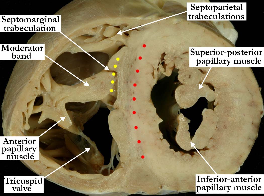
Modality: Anatomic specimen
Orientation: Mid-ventricular short axis view
Description: The section is from the same
heart as shown in image
A010100-141a and
A010100-142a, but is taken closer yet to the base of the heart. This
short axis view demonstrates similar features to those shown in image
A010100-142a, but
with subtle changes in the anatomy along the ventricular septum. At the
level of the tips of the papillary muscles within the left ventricle, the
interventricular septum begins to have a smooth appearance, and the aortic,
or anterior, leaflet of the mitral valve is lifted away from the ventricular
septum. In contrast, the right ventricular component of the interventricular
septum remains coarsely trabeculated, with the septal leaflet of the
tricuspid valve lying immediately adjacent to the septum. The body of the
septomarginal trabeculation is better seen at this level (yellow dots),
adjacent with the moderator band as it branches from this prominent septal
structure of the right ventricleThe muscular interventricular septum is
marked with red dots.
Contributor: Diane
E. Spicer, BS
Institution: The Congenital Heart
Institute of Florida (CHIF)
Image Label: A010100-143a
Source of Image: The Congenital Heart
Institute of Florida (CHIF)
Image Certification: pending
AWG Rating: pending
|