|
Derived Terms: |
|
|
AEPC: |
Normal heart (01.01.00) |
| |
Normal position-orientation of heart (02.01.00) |
| |
|
|
EACTS-STS: |
Normal heart (01.01.00) |
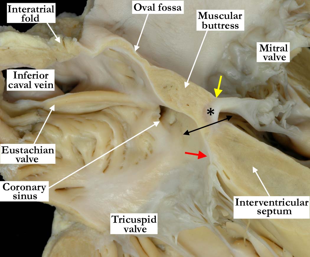
Modality: Anatomic specimen
Orientation: Four chamber view
Description: This simulated four chamber
echocardiographic view is taken from the same heart as shown in image
A010100-137a,
but is sectioned more posteriorly and inferiorly. The cut demonstrates the
overlapping areas of atrial and muscular myocardium, which produce the
so-called atrioventricular muscular sandwich. The atrial muscular buttress
is just beginning to overlap the crest of the muscular ventricular septum.
The fibrous tissue at the apex of the triangle of Koch is along the postero-inferior
component of the aortic root (black asterisk). In this section, the
atrioventricular component of the mebranpuis septum forms part of the normal
offseting of the hinges of the atrioventricular valves. The tricuspid valve
(red arrow) is in a more apical position with respect to the muscular
interventricular septum. The yellow arrow marks the superior insertion of
the mitral valve.
Contributor: Diane
E. Spicer, BS
Institution: The Congenital Heart
Institute of Florida (CHIF)
Image Label: A010100-138a
Source of Image:
Idriss Archive, Lurie Childrens Hospital,
Chicago, IL
Image Certification: pending
AWG Rating: pending
|