|
Derived Terms: |
|
|
AEPC: |
Normal heart (01.01.00) |
| |
Normal position-orientation of heart (02.01.00) |
| |
|
|
EACTS-STS: |
Normal heart (01.01.00) |
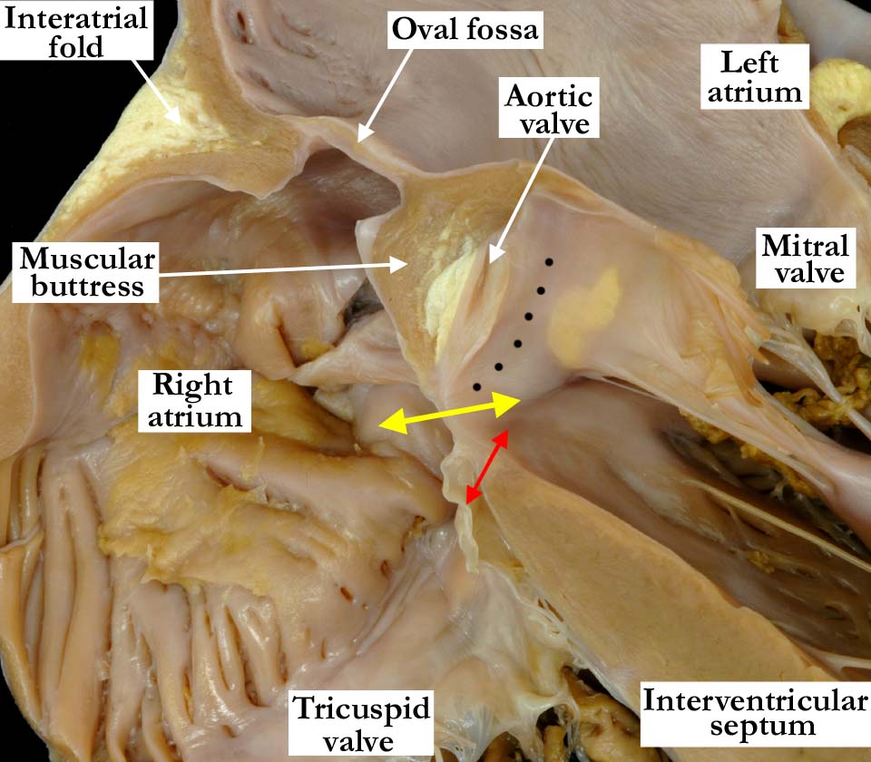
Modality: Anatomic specimen
Orientation: Four chamber view
Description: The heart is sectioned to
simulate a four chamber echocardiographic view, with the section cut towards
the posterior inlet portion of the ventricles. The tricuspid valve separates
the membranous septum into atrioventricular (yellow arrow) and
interventricular (red arrow) portions. Note the extensive area of fibrous
continuity between the aortic and mitral valves (black dots). Please
click on
A010100-138a to view the companion image for this heart.
Contributor: Diane
E. Spicer, BS
Institution: The Congenital Heart
Institute of Florida (CHIF)
Image Label: A010100-137a
Source of Image:
Idriss Archive, Lurie Childrens Hospital,
Chicago, IL
Image Certification: pending
AWG Rating: pending
|