|
||||||||
|
|
|||||||||
|
|||||||||
|
IPCCC Code:
06.01.03, 07.02.00, 07.05.30 |
|||
|
AEPC Derived Term: |
Tricuspid valvar dysplasia (06.01.03) Right ventricular hypoplasia (07.02.00) Pulmonary stenosis, subvalvar (07.05.30) |
||
|
EACTS-STS Derived Term: |
Tricuspid valve disease, Tricuspid valve pathology, Mucoid thickening
(Tricuspid valve dysplasia) (06.01.03) Hypoplastic right ventricle (07.02.00) Pulmonary stenosis, subvalvar (07.05.30) |
||
|
ICD10 Derived Term: |
Other congenital malformations of tricuspid valve (Q22.8) Other congenital malformations of cardiac chambers and connections (Q20.8) |
||
|
Definition: NA |
|
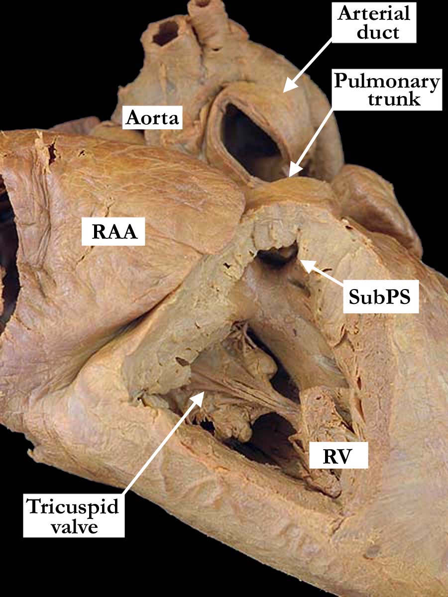
Modality: Anatomic specimen Orientation: Right heart view Description: In this anatomical view of the heart, the right atrial appendage (RAA) is dilated. Although the right ventricle (RV) is hypoplastic, the right ventricular hypoplasia is not well demonstrated in this image, because one cannot see the right ventricular apex. The window cut into the right ventricular wall shows the thickened, dysplastic tricuspid valve in the inlet portion. The right ventricle is hypertrophied and there is subpulmonary/infundibular pulmonary stenosis (SubPS). Contributor: Diane Spicer, BS Institution: The Congenital Heart Institute of Florida (CHIF) Image Label: A090501-32a Source of Image: Van Mierop Archive, University of Florida, Gainesville, Florida Image Certification: 10 May 2014
AWG Rating:
|
|
|||
|
This gross morphologic specimen has also been presented in a separate web page focused on the combined malformations of both semilunar valves. Please click here to view this additional web page. |
||||
AWG Certification: 10 May 2014
|
Copyright ipccc-awg.net All Rights Reserved. Frontpage-Templates.org |