|
||||||||
|
|
|||||||||
|
|||||||||
|
IPCCC:
09.15.22, 09.05.32, 01.03.10 |
|||
|
AEPC Derived Term: |
Bicuspid aortic
valve (09.15.22) |
||
|
EACTS-STS Derived Term: |
Aortic valve
pathology, Bicuspid (09.15.22) |
||
|
ICD10 Derived Term: |
Congenital stenosis of aortic
valve (Q23.0) |
||
|
Definition: pending
Comments: This series of images from the same specimen illustrate the use of the the interleaflet triangles as a means of recognizing the overall nature of valves with two leaflets, and also those with only one leaflet (the so-called unicommissural and unicuspid variant). These images illustrate the bifoliate nature of the pulmonary and aortic valves of this specimen. Please note that the presence of three attachments at the sinotubular junction does not mean that the valve is trifoliate. The number of leaflets should be determined on the pattern of opening and closing of the valve, and these valves can only open and close in bifoliate fashion, since it has a solitary zone of apposition within the skirt of leaflet tissue. It is usual to find interleaflet triangles in the setting of bifoliate valves but only rarely is the arrangement both bifoliate and bisinuate! Thus, the presence of the interleaflet triangle does not disqualify the valve from being bifoliate. For further information on the subject please refer to these references:
|
|
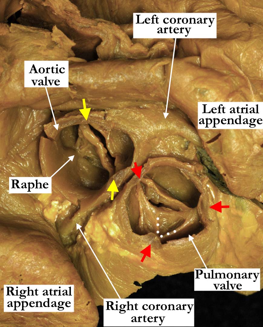
Modality: Anatomic specimen Orientation: Short axis view (base) Description: This short axis view of the base of the heart shows the aortic and pulmonary valves. The aortic valve is bicuspid, with the attachments of the leaflets at the sinotubular junction marked with the yellow arrows. The right and left coronary arteries arise from separate sinuses. A raphe is present within the aortic sinus giving rise to the right coronary artery. The pulmonary valve is similarly bifoliate, and the valve is markedly stenotic. The white dots illustrate an area of fusion between two primordiums initially present during development, with the presence of an interleaflet triangle on the ventricular aspect supporting the notion of fusion during development Contributor: Diane E. Spicer, BS Institution: The Congenital Heart Institute of Florida (CHIF) Image Label: A091522-22a Source of Image: Van Mierop Archive, University of Florida, Gainesville, Florida Image Certification: 5 May 2012
AWG Rating:
|
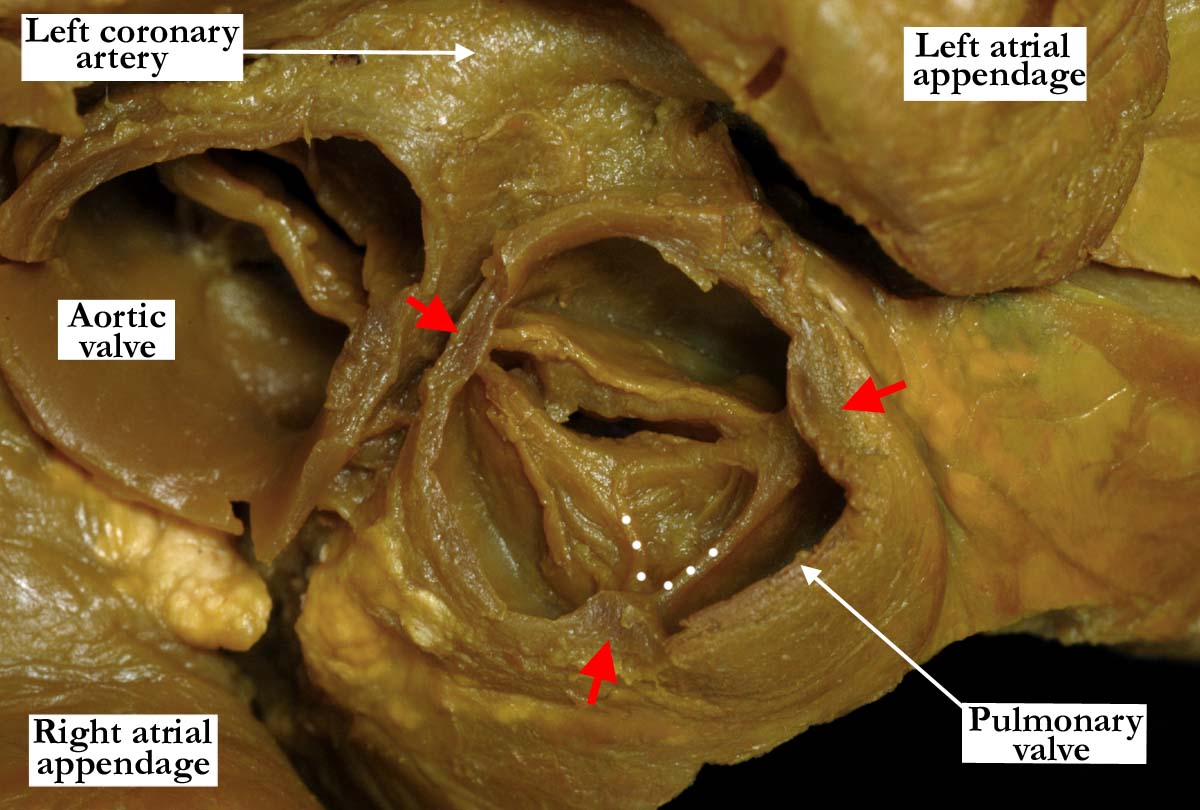
Modality: Anatomic specimen Orientation: Short axis view (base) Description: This short axis view focuses on the pulmonary valve. There is but a solitary zone of apposition, pointing to the presence of two leaflets, but the arrangement at the sinotubular junction, with three peripheral attachments marked with red arrows, indicates that the valve started development with three putative leaflets. The valve is markedly stenotic, with a slit-like opening. The presence of three interleaflet triangles (see image A091522-22c) on the ventricular aspect and the solitary zone of apposition, lend further support to the interpretation of the valve being bifoliate, with fusion of the anterior-leftward and anterior-rightward leaflets during development. Contributor: Diane E. Spicer, BS Institution: The Congenital Heart Institute of Florida (CHIF) Image Label: A091522-22b Source of Image: Van Mierop Archive, University of Florida, Gainesville, Florida Image Certification: 5 May 2012
AWG Rating:
|
|||
|
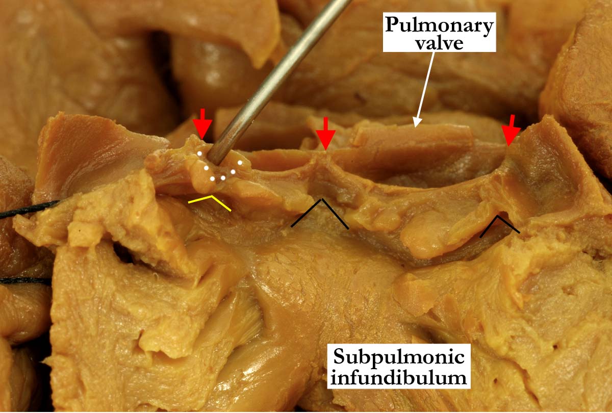
Modality: Anatomic specimen Orientation: Opened right ventricular outflow tract view Description: The stenotic pulmonary valve is opened, demonstrating the thickened, nodular edges of the valve, along with the fused area (white dots) between two of the leaflets. The fused leaflets have been lifted to demonstrate the hypoplastic interleaflet triangle seen on the ventricular aspect (yellow lines). Note that this triangle is less well formed than the remaining two interleaflet triangles (black lines). The red arrows mark the attachment of the leaflets at the sinotubular junction. Contributor: Diane E. Spicer, BS Institution: The Congenital Heart Institute of Florida (CHIF) Image Label: A091522-22c Source of Image: Van Mierop Archive, University of Florida, Gainesville, Florida Image Certification: 5 May 2012
AWG Rating:
|
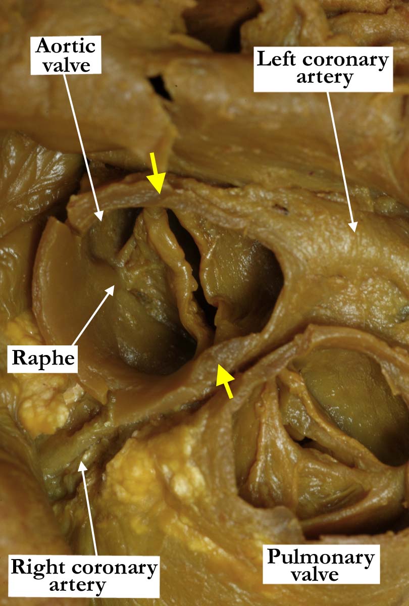
Modality: Anatomic specimen Orientation: Short axis view (base) Description: This short axis view focuses on the aortic valve. The aortic valve is bicuspid, with the attachments at the sinutubular junction marked with the yellow arrows. The valve is thickened, with a small raphe within the right-sided coronary sinus, made up of the fused right coronary and non-coronary aortic sinuses. The right and left coronary arteries exit the aorta from separate sinuses adjacent to the pulmonary trunk. Contributor: Diane E. Spicer, BS Institution: The Congenital Heart Institute of Florida (CHIF) Image Label: A091522-22d Source of Image: Van Mierop Archive, University of Florida, Gainesville, Florida Image Certification: 5 May 2012
AWG Rating:
|
|||
|
Modality: Anatomic specimen Orientation: Opened left ventricular outflow tract view Description: The aortic valve has been opened, the cut edges of the left coronary leaflet marked with black dots. The two attachments at the sinutubular junction are marked with yellow arrows. There are two well-formed interleaflet triangles (black lines) on the ventricular aspect of the valve, along with an hypoplastic triangle beneath the zone of fusion between the initial right coronary and non-coronary aortic leaflets (red arrow). Contributor: Diane E. Spicer, BS Institution: The Congenital Heart Institute of Florida (CHIF) Image Label: A091522-22e Source of Image: Van Mierop Archive, University of Florida, Gainesville, Florida Image Certification: 5 May 2012
AWG Rating:
|
|
|||
|
This gross morphologic specimen has also been presented in a separate web page focused on the malformations of the tricuspid valve and right ventricle. Please click here to view this additional web page. |
||||
AWG Page Certification: 5 May 2012
|
Copyright ipccc-awg.net All Rights Reserved. Frontpage-Templates.org |