|
|||||
|
|
||||||
|
||||||
|
IPCCC: 07.08.50, 03.01.05 [01.03.03], 01.04.01, 01.05.01 |
|||
|
AEPC Derived Term: |
Ventricular myocardial noncompaction cardiomyopathy (07.08.50) Left isomerism ('polysplenia') (03.01.05) vs Isomerism of left atrial appendages (left isomerism) (01.03.03) Discordant AV
connections
(01.04.01) |
||
|
EACTS-STS Derived Term: |
Cardiomyopathy, Spongiform cardiomyopathy (Noncompaction cardiomyopathy) (Embryonal cardiomyopathy) (07.08.50) Atrial appendage isomerism, Left (03.01.05) vs Syndrome, Heterotaxy (heterotaxy syndrome) (visceral heterotaxy)-modifier, Isomerism, Isomerism of the left atrial appendages AV connection(s) = Discordant AV connections (biventricular) (01.04.01) VA connection =Discordant VA connections (TGA) (01.05.01) |
||
|
Definition: pending
|
|
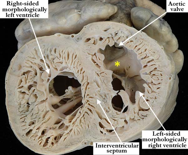
Modality: Anatomic specimen Orientation: Short axis - apical view Description: This apical view of the ventricles demonstrates biventricular, spongiform myocardium or so-called non-compaction. There is a subendocardial, compact zone with a highly trabeculated inner myocardial component. The atrioventricular connections are discordant in this heart with isomerism of the left atrial appendages, ventricular inversion and left-hand ventricular topology. There are discordant ventriculo-arterial connections, the aorta arising from the left-sided morphologically right ventricle above a subaortic infundibulum (yellow asterisk). Contributor: Diane E. Spicer, BS Institution: The Congenital Heart Institute of Florida (CHIF) Image Label: A070850-80a Source of Image: Van Mierop Archive, University of Florida Image Certification: pending AWG Rating: pending
|
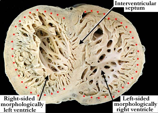
Modality: Anatomic specimen Orientation: Short axis - apical view Description: This view illustrates the apical trabecular pattern of both ventricles allowing for morphologic differentiation in this heart with spongiform myocardium. The left-sided, coarsely trabeculated ventricle is consistent with a morphologically right ventricle, while the right-sided ventricle has fine apical trabeculations, consistent with a morphologically left ventricle. The subendocardial compact zone is shown between the epicardium and the red dots, the highly trabeculated myocardium closely related to the ventricular cavity and more prominent within the left ventricle. Contributor: Diane E. Spicer, BS Institution: The Congenital Heart Institute of Florida (CHIF) Image Label: A070850-80b Source of Image: Van Mierop Archive, University of Florida Image Certification: pending AWG Rating: pending |
|||
| click here for additional images associated with this heart |
AWG Page Certification: pending
|
Copyright ipccc-awg.net All Rights Reserved. Frontpage-Templates.org |