|
|||||
|
|
||||||
|
||||||
|
IPCCC: 03.01.05 [01.03.03], 01.04.01, 01.05.01, 07.08.50 |
|||
|
AEPC Derived Term: |
Left isomerism ('polysplenia') (03.01.05) vs Isomerism of left atrial appendages (left isomerism) (01.03.03) Discordant AV
connections
(01.04.01) Ventricular myocardial noncompaction cardiomyopathy (07.08.50) |
||
|
EACTS-STS Derived Term: |
Atrial appendage isomerism, Left (03.01.05) vs Syndrome, Heterotaxy (heterotaxy syndrome) (visceral heterotaxy)-modifier, Isomerism, Isomerism of the left atrial appendages AV connection(s) = Discordant AV connections (biventricular) (01.04.01) VA connection =Discordant VA connections (TGA) (01.05.01) Cardiomyopathy, Spongiform cardiomyopathy (Noncompaction cardiomyopathy) (Embryonal cardiomyopathy) (07.08.50) |
||
|
Definition: pending
|
|
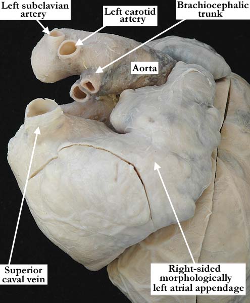
Modality: Anatomic specimen Orientation: Lateral view Description: This lateral view of the right-sided, morphologically left atrial appendage demonstrates the characteristic, constricted junction of the atrial appendage to the atrial vestibule. As well, the appendage is narrow with scalloped edges. The superior caval vein is right-sided. The aorta arches to the left with normal branching of the brachiocephalic vessels. Contributor: Diane E. Spicer, BS Institution: The Congenital Heart Institute of Florida (CHIF) Image Label: A030105-81a Source of Image: Van Mierop Archive, University of Florida Image Certification: pending AWG Rating: pending
|
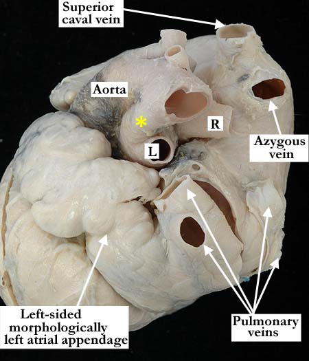
Modality: Anatomic specimen Orientation: Posterior lateral view Description: This posterior, lateral view of the left-sided, morphologically left atrial appendage demonstrates the characteristic, constricted or narrow junction of the atrial appendage to the atrial vestibule. The left-sided appendage itself is narrow with scalloped edges, this more so than in the right-sided appendage shown in UF86-115c. The pulmonary veins drain to the left-sided atrium. Note the large caliber of the azygous vein where it exits the right-sided superior caval vein. The aorta exits the ventricular mass anterior to the pulmonary trunk. The left and right pulmonary arteries bifurcate from the pulmonary trunk in the usual fashion and there is a left-sided, patent arterial duct (yellow asterisk). Contributor: Diane E. Spicer, BS Institution: The Congenital Heart Institute of Florida (CHIF) Image Label: A030105-81b Source of Image: Van Mierop Archive, University of Florida Image Certification: pending AWG Rating: pending
|
|||
|
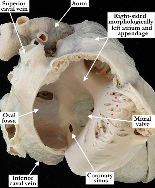
Modality: Anatomic specimen Orientation: Lateral view Description: The right-sided, morphologically left atrium has been windowed and its internal structure is viewed from the lateral aspect. The pectinate muscles are confined to the atrial appendage with some spilling out onto the anterior, lateral wall of the atrium, with a smooth-walled vestibule at the atrioventricular junction. The pectinate muscles do not extend to the crux of the heart. There is a coronary sinus and a small atrial septal defect within the oval fossa. The superior and inferior caval veins are right-sided. The mitral valve guards the inlet to the right-sided, morphologically left ventricle. Contributor: Diane E. Spicer, BS Institution: The Congenital Heart Institute of Florida (CHIF) Image Label: A030105-81c Source of Image: Van Mierop Archive, University of Florida Image Certification: pending AWG Rating: pending
|
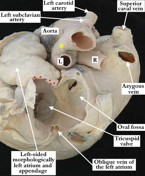
Modality: Anatomic specimen Orientation: Posterior lateral view Description: The left-sided, morphologically left atrium has been windowed and is viewed from the posterior, lateral aspect. The atrial vestibule is smooth and the pectinate muscles (red dots) are confined to the atrial appendage. The left aspect of the oval fossa and the small atrial septal defect are easily appreciated. The oblique vein of the left atrium lies within the posterior, inferior wall of the left-sided atrium as it extends from the coronary sinus toward the left atrioventricular groove. The tricuspid valve guards the inlet. Contributor: Diane E. Spicer, BS Institution: The Congenital Heart Institute of Florida (CHIF) Image Label: A030105-81d Source of Image: Van Mierop Archive, University of Florida Image Certification: pending AWG Rating: pending |
|||
| click here for additional images associated with this heart |
AWG Page Certification: pending
|
Copyright ipccc-awg.net All Rights Reserved. Frontpage-Templates.org |