|
||||||||
|
|
|||||||||
|
|||||||||
|
IPCCC: 09.15.22 |
|||
|
AEPC Derived Term: |
Bicuspid aortic valve (09.15.22) |
||
|
EACTS-STS Derived Term: |
Aortic valve pathology, Bicuspid (09.15.22) |
||
|
ICD10 Derived Term: |
Congenital insufficiency of aortic valve: Bicuspid aortic valve (Q23.1) | ||
|
Definition: pending |
|
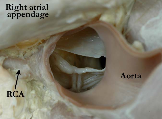
Modality: Anatomic specimen Orientation: Arterial view of the aorta Description: This arterial view of the aortic valve demonstrates the bifoliate nature of the valve. The right and left coronary leaflets are fused with a prominent raphe demonstrating the line of non-separation between the conjoined leaflets. The edge of the conjoined leaflets is thickened, rendering the valve insufficient. The non-coronary leaflet is of normal thickness. (RCA - right coronary artery). Contributor: Diane Spicer, BS Institution: Congenital Heart Institute of Florida Image Label: A091522-60a Source of Image: Van Mierop Archive, University of Florida, Gainesville, Florida Image Certification: pending AWG Rating: pending
|
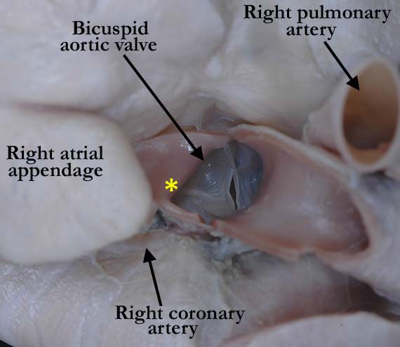
Modality: Anatomic specimen Orientation: Arterial view of the aorta Description: The right and non-coronary leaflets are fused in this bifoliate aortic valve. Note the prominent raphe (*) showing the line of non-separation between the conjoined leaflets. Contributor: Diane Spicer, BS Institution: Congenital Heart Institute of Florida Image Label: A091522-60b Source of Image: Van Mierop Archive, University of Florida, Gainesville, Florida Image Certification: pending AWG Rating: pending |
|||
|
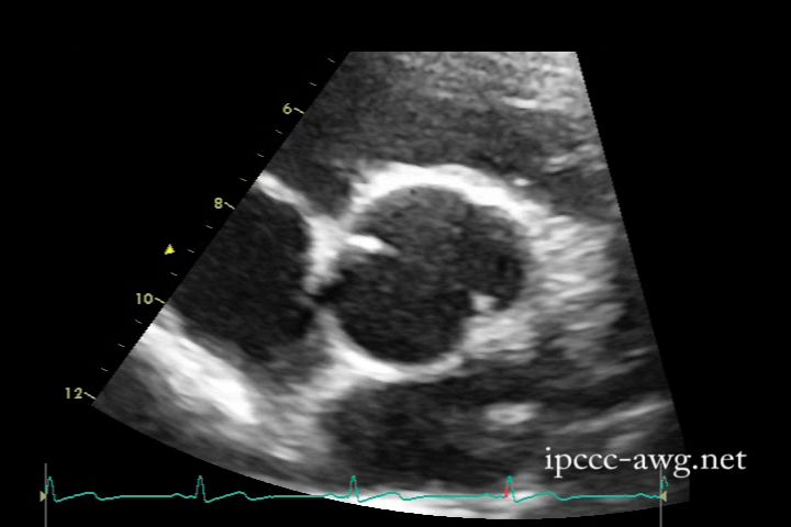
Modality: Transthoracic 2D echocardiogram Orientation: Cross sectional view of the aorta Description: This image shows a bicuspid aortic valve formed by the fusion of the right and left coronary cusps. Contributor: Douglas Sexton Institution: Congenital Heart Institute of Florida Image Label: E091522-60c Source of Image: Congenital Heart Institute of Florida Image Certification: pending AWG Rating: pending
|
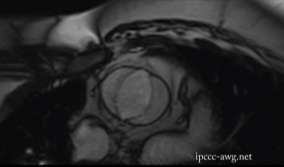
Modality: Cardiac MRI image Orientation: Short axis Description: MR cine SSFP short-axis image of the bicuspid aortic valve due to the fusion of the left and noncoronary leaflets. Contributor: Charles W. Shepard, MD Institution: University of Minnesota Amplatz Childrens Hospital Image Label: MR091522-60d Image Source: Heart Center, University of Minnesota Amplatz Childrens Hospital Minneapolis, Minnesota Image Certification: pending AWG Rating: pending |
|||
|
For an excellent commentary on the anatomical perspectives of using the interleaflet triangles (as a means of recognizing the overall nature of semilunar valves with two leaflets) please click here |
||||
AWG Page Certification: pending
|
Copyright ipccc-awg.net All Rights Reserved. Frontpage-Templates.org |