|
||||||||
|
|
|||||||||
|
|||||||||
|
IPCCC: 14.10.42, 10.10.12, 09.15.06, 06.02.42 |
|||
|
AEPC Derived Term: |
Fetal closure of
oval foramen (14.10.42) Aortic valvar atresia (09.15.06) Arcade abnormality of mitral chords-papillary muscles (06.02.42) |
||
|
EACTS-STS Derived Term: |
Fetal diagnosis,
Closed foramen ovale (14.10.42) Aortic valve atresia (09.15.06) Mitral valve disease, Mitral valve pathology, Mitral chordal abnormality, Arcade abnormality of mitral chords/ papillary muscles (06.02.42) |
||
|
ICD10 Derived Term: |
Abnormal ultrasonic finding on antenatal screening of mother (O28.3) Endocardial fibroelastosis (I42.4) Congenital aortic stenosis or atresia (Q23.0) Other specified congenital malformations of heart (Q24.8) |
||
|
Definition: pending
|
|
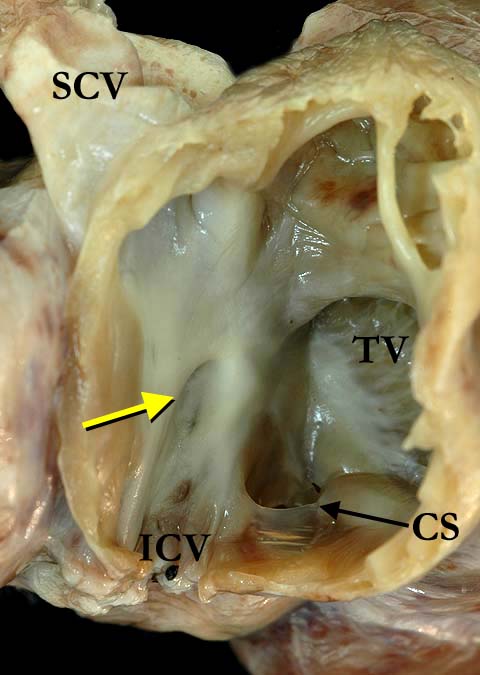
Modality: Anatomic specimen Orientation: Anterior oblique right atrial view Description: A window cut in the anterior lateral wall of the right atrium shows the atrial septum, the septal portion of the tricuspid valve (TV) and the coronary sinus (CS). The superior (SCV) and inferior (ICV) caval veins connect to the right atrium in the usual fashion. The arrow points to a small oval fossa on the atrial septal surface. The oval foramen (foramen ovale), which was expected to be at the antero-superior margin of the oval fossa, was prematurely closed in this case. Note the endocardial fibroelastosis within the right atrium. This case was diagnosed in utero with critical aortic stenosis that eventually progressed to aortic atresia. Contributor: Diane E. Spicer, BS Institution: The Congenital Heart Institute of Florida (CHIF) Image Label: A050302-10a Source of Image: The Congenital Heart Institute of Florida (CHIF) Image Certification: 5 August 2011
AWG Rating:
|
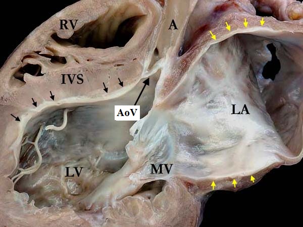
Modality: Anatomic specimen Orientation: Long axis view Description: This long axis view demonstrates a dilated, markedly thickened left atrium (LA) in a heart with premature closure of the oval foramen (foramen ovale). There is endocardial thickening of both the atrium (yellow arrows) and the ventricle (black arrows). The mitral valve (MV) is thickened and attached directly to the tips of the papillary muscle (mitral arcade). The aortic valve (AoV) is imperforate. (IVS-interventricular septum, LV-left ventricle, RV-right ventricle) Contributor: Diane E. Spicer, BS Institution: The Congenital Heart Institute of Florida (CHIF) Image Label: A050302-10b Source of Image: The Congenital Heart Institute of Florida (CHIF) Image Certification: 5 August 2011
AWG Rating:
|
|||
|
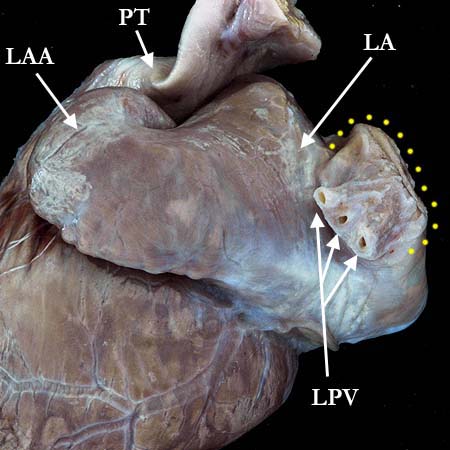
Modality: Anatomic specimen Orientation: Posterior left atrial view Description: The left atrial appendage (LAA) and the left atrium (LA) are markedly dilated. The left pulmonary veins (LPV) enter the left atrium via a dilated common vein that is highlighted with yellow dots. (PT-pulmonary trunk). Contributor: Diane E. Spicer, BS Institution: The Congenital Heart Institute of Florida (CHIF) Image Label: A050302-10c Source of Image: The Congenital Heart Institute of Florida (CHIF) Image Certification: 5 August 2011
AWG Rating:
|
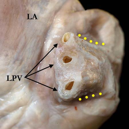
Modality: Anatomic specimen Orientation: Posterior left atrial view (close up) Description: In the close up view, the left pulmonary veins (LPV) demonstrate thickening of the venous walls and intimal surface. The dilated area where the three pulmonary veins enter the left atrium (LA) is marked with the yellow dots. Contributor: Diane E. Spicer, BS Institution: The Congenital Heart Institute of Florida (CHIF) Image Label: A050302-10d Source of Image: The Congenital Heart Institute of Florida (CHIF) Image Certification: 5 August 2011
AWG Rating:
|
|||
AWG Page Certification: 5 August 2011
|
Copyright ipccc-awg.net All Rights Reserved. Frontpage-Templates.org |