|
|||||
|
|
||||||
|
||||||
|
IPCCC: 09.28.37 |
|||
|
AEPC Derived Term: |
Double aortic arch with right arch dominant: left arch atretic (09.28.37) | ||
|
EACTS-STS Derived Term: |
Vascular ring, Double aortic arch, Right arch dominant, Left arch atretic (09.28.37) | ||
|
Definition: pending |
|
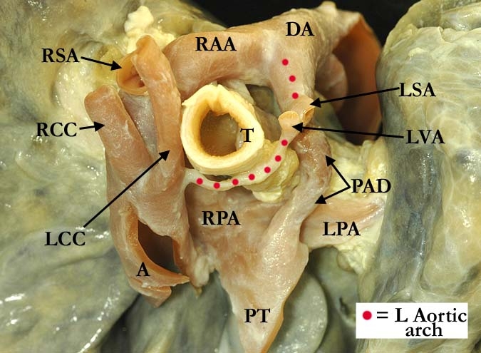
Modality: Anatomic specimen Orientation: Cephalo-caudal Description: Viewed from above is a vascular ring that is composed of a double aortic arch with atresia of the left aortic arch (red dots). The right aortic arch (RAA) is dominant with the left common carotid (LCC) and right common carotid (RCC) arteries having a near common origin as the first branches from the right arch. The right subclavian artery (RSA) branches from the right arch distal to the carotid arteries. The distal portion of the left aortic arch is patent as it attaches to the descending aorta (DA), this portion of the arch typically referred to as the diverticulum of Kommerell. The left subclavian artery (LSA) and left vertebral artery (LVA) branch from this diverticulum, distal to the atretic segment of the left aortic arch. The patent arterial duct (PAD) branches from the patent portion of the left aortic arch, forming an additional U-shaped ring around the trachea (T). The esophagus was previously removed from its anatomic position. (A-aorta, PT-pulmonary trunk, LPA-left pulmonary artery, RPA- right pulmonary artery) Contributor: Diane Spicer, BS Institution: The Congenital Heart Institute of Florida (CHIF) Image Label: A092837-66a Source of Image: Idriss Archive, Childrens Memorial Hospital, Chicago, IL Image Certification: pending AWG Rating: pending
|
|||
AWG Page Certification: pending
|
Copyright ipccc-awg.net All Rights Reserved. Frontpage-Templates.org |