|
|||||
|
|
||||||
|
||||||
|
IPCCC: 01.05.01, 07.11.01, 09.29.01, 09.29.12, 06.01.09
or 01.05.01, 07.10.00, 09.28.20 |
|||
|
AEPC Derived Term: |
Discordant VA connections (TGA) (01.05.01) Muscular VSD (07.11.01) Aortic coarctation (09.29.01) Aortic arch hypoplasia (tubular) distal to subclavian artery (isthmal) (09.29.12) Straddling tricuspid valve (06.01.09) |
||
|
EACTS-STS Derived Term: |
TGA - VSD - Aortic arch narrowing (Transposition
of the Great Arteries - Ventricular septal defect - Aortic arch narrowing)
(Concordant atrioventricular connections and Discordant ventriculo-arterial
connections - VSD - Aortic arch narrowing)(01.05.01, 07.10.00, 09.28.20) VSD, Type 4 (Muscular)(07.11.01) Coarctation of the Aorta (CoAo)-modifier, With isthmus hypoplasia (distal to the subclavian artery)(09.29.01, 09.29.12) Tricuspid valve disease, Tricuspid valve pathology, Straddling tricuspid valve (06.01.09) |
||
|
Definition: pending |
|
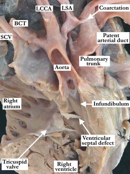
Modality: Anatomic specimen Orientation: Anterior superior view Description: The free wall of the right atrium and right ventricle, along with the anterior aspect of the aorta have been removed in this heart with transposition of the great arteries. The pulmonary trunk is posterior and to the left of the aorta. The aorta is stenotic and is supported by a complete muscular infundibulum. Distally, there is a severe aortic coarctation with narrowing of the isthmus between the left common carotid artery (LCCA) and the patent arterial duct. There are concordant atrioventricular connections and a non-committed, muscular ventricular septal defect. (SCV-superior caval vein, BCT-brachiocephalic trunk, LSA-left subclavian artery) Contributor: Diane Spicer, BS Institution: The Congenital Heart Institute of Florida (CHIF) Image Label: A010501-71a Source of Image: The Congenital Heart Institute of Florida (CHIF) Image Certification: pending AWG Rating: pending
|
Modality: Anatomic specimen Orientation: Anterior superior view Description: A close up view of the opened, patent arterial duct and descending aorta shown in the left panel demonstrates the extremely stenotic aortic isthmus where it joins the descending aorta. Note that this orifice is surrounded by the wrinkled intima that extends from the patent arterial duct. (BT-brachiocephalic trunk, LCC-left common carotid artery, LSA-left subclavian artery) Contributor: Diane Spicer, BS Institution: The Congenital Heart Institute of Florida (CHIF) Image Label: A010501-71b Source of Image: The Congenital Heart Institute of Florida (CHIF) Image Certification: pending AWG Rating: pending |
|||
|
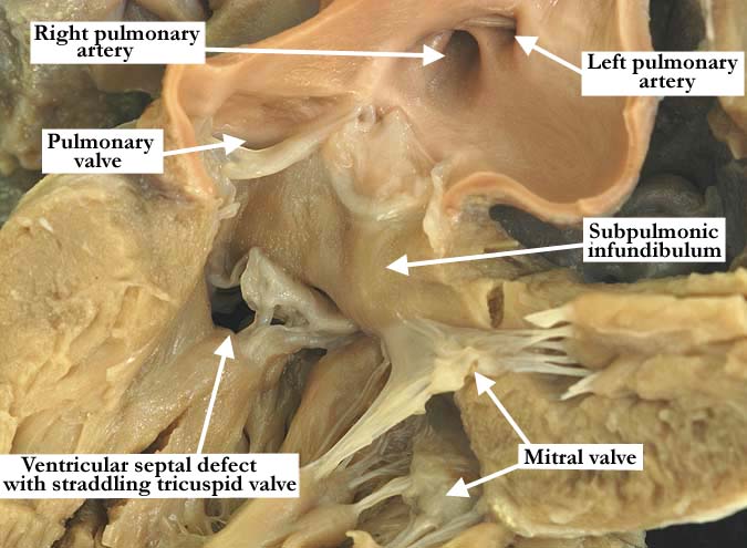
Modality: Anatomic specimen Orientation: Left ventricular view Description: This close up view of the same heart shown in above shows the outlet component of this morphologically left ventricle in this heart with discordant ventriculo-arterial connections and concordant atrioventricular connections (transposition of the great arteries). The pulmonary trunk is supported by a complete muscular infundibulum as it exits the left ventricle. There is absence of fibrous continuity between the arterial and atrioventricular valves. The tricuspid valve straddles the interventricular septum through a muscular ventricular septal defect. Contributor: Diane Spicer, BS Institution: The Congenital Heart Institute of Florida (CHIF) Image Label: A010501-71c Source of Image: The Congenital Heart Institute of Florida (CHIF) Image Certification: pending AWG Rating: pending
|
AWG Page Certification: pending
|
Copyright ipccc-awg.net All Rights Reserved. Frontpage-Templates.org |