|
Derived Terms: |
|
|
AEPC: |
Normal heart (01.01.00) |
| |
Normal position-orientation of heart (02.01.00) |
| |
|
|
EACTS-STS: |
Normal heart (01.01.00) |
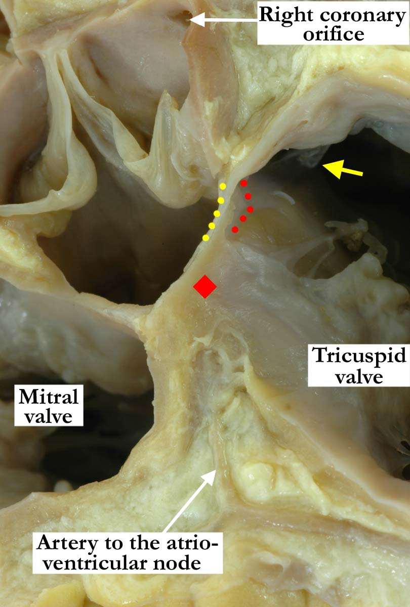
Modality: Anatomic specimen
Orientation: Short axis view base of the
heart
Description: Having removed the atrial
myocardium forming the atrial surface of the triangle of Koch, and with the
floor of the coronary sinus dissected away, the artery to the
atrioventricular node is revealed. This artery lies in a paraseptal space
between the atrial musculature and the crest of the muscular ventricular
septum. The artery extends anteriorly to the apex of the triangle of Koch
(red diamond). The non-coronary leaflet of the aortic valve and a portion of
the septal leaflet of the tricuspid valve have also been removed, revealing
the atrioventricular (yellow dots) and interventricular components of the
membranous septum (red dots). Note the close relationship of the aortic
valve, the membranous septum, and the tricuspid valve. The medial papillary
muscle is marked with a yellow arrow.
Contributor: Diane
E. Spicer, BS
Institution: The Congenital Heart
Institute of Florida (CHIF)
Image Label: A010100-136a
Source of Image: The Congenital Heart
Institute of Florida (CHIF)
Image Certification: pending
AWG Rating: pending
|