|
Derived Terms: |
|
|
AEPC: |
Normal heart (01.01.00) |
| |
Normal position-orientation of heart (02.01.00) |
| |
|
|
EACTS-STS: |
Normal heart (01.01.00) |
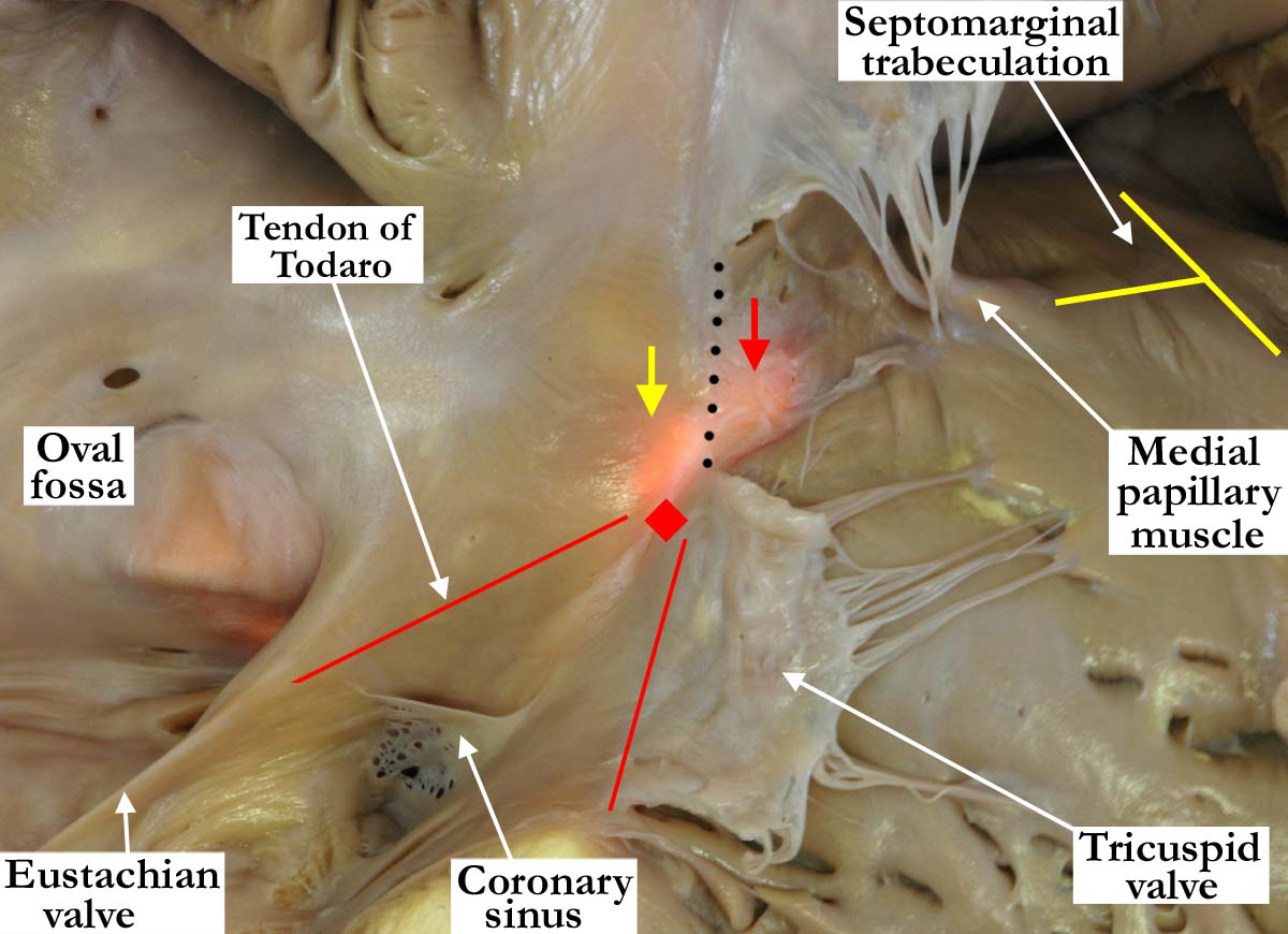
Modality: Anatomic specimen
Orientation:
Anterior,
right ventricular view
Description: A portion of the septal
leaflet of the tricuspid valve has been removed in this close up view of the
right atrioventricular junction. A light placed behind the membranous septum
demonstrates both the atrioventricular (yellow arrow) and interventricular
(red arrow) components, the black line marking where the tricuspid valve
divides the two portions. The atrioventricular node (red diamond) is usually
located within the musculature of the antero-inferior buttress of the atrial
septum at the apex of the triangle of Koch. The triangle of Koch itself is
delineated by the tendon of Todaro, which is a continuation of the
Eustachian valve, and the hinge line of the septal leaflet of the tricuspid
valve. The coronary sinus lies at the base of the triangle. The
septomarginal trabeculation (yellow Y) divided at the ventricular base
into two limbs, with the supraventricular crest inserting between the limbs.
The medial papillary muscle arises from the postero-caudal limb, while the
antero-cephalad limb extends to to support the leftward adjacent leaflet of
the pulmonary valve. For a left ventricular view of this heart, please
see companion image
A010100-135a.
Contributor: Diane
E. Spicer, BS
Institution: The Congenital Heart
Institute of Florida (CHIF)
Image Label: A010100-134a
Source of Image: The Congenital Heart
Institute of Florida (CHIF)
Image Certification: pending
AWG Rating: pending
|