|
|||||
|
|
||||||
|
||||||
|
IPCCC: AEPC: 09.15.03, 06.02.92, 10.10.12, 09.45.12, 12.10.19 EACTS-STS: 01.01.09, 09.15.03, 06.02.92, 07.21.00, 01.01.09, 09.45.00, Q1.45.52,
01.01.09, 10.10.12, 12.10.01, 12.31.06,
12.10.00 |
|||
|
AEPC Derived Term: |
Hypoplastic left heart syndrome, aortic atresia (09.15.03),
mitral stenosis (06.02.92), Endocardial fibroelastosis (10.10.12), Coronary fistulas from left ventricle ('sinusoidal') (09.45.12), Norwood type procedure: neoaortic construction with interposition conduit between pulmonary trunk + aortic arch (12.10.19) |
||
|
EACTS-STS Derived Term: |
Hypoplastic left heart syndrome (HLHS), Aortic atresia +
Mitral stenosis, IVS (01.01.09, 09.15.03, 06.02.92, 07.21.00), Hypoplastic left heart syndrome (HLHS)-modifier-Coronary artery fistula(s) or sinusoid(s), (Coronary-cameral fistula(s)) present-Involved coronary = LAD (01.01.09, 09.45.00, Q1.45.52), Hypoplastic left heart syndrome (HLHS)-modifier, Endocardial fibroelastosis (01.01.09, 10.10.12), Norwood (Stage 1), Neoaortic construction using arch augmentation with patch (12.10.01), Norwood (Stage 1)- modifier, Source of pulmonary blood flow, Shunt - systemic artery to pulmonary artery (12.31.06, 12.10.00) |
||
|
Definition: pending |
|
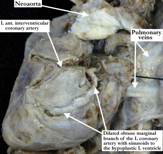
Modality: Anatomic specimen Orientation: Surface view of the left ventricle Description: This hypoplastic left heart is status post Norwood procedure. The left anterior interventricular branch is small and lies within the muscle along the majority of its course. There is a markedly dilated obtuse marginal branch that dives into the muscle toward the apex of the hypoplastic left ventricle. The neoaorta and the pulmonary veins provide anatomic orientation. Contributor: Diane Spicer, BS Institution: The Congenital Heart Institute of Florida (CHIF) Image Label: A091503-66a Image Certification: pending AWG Rating: pending
|
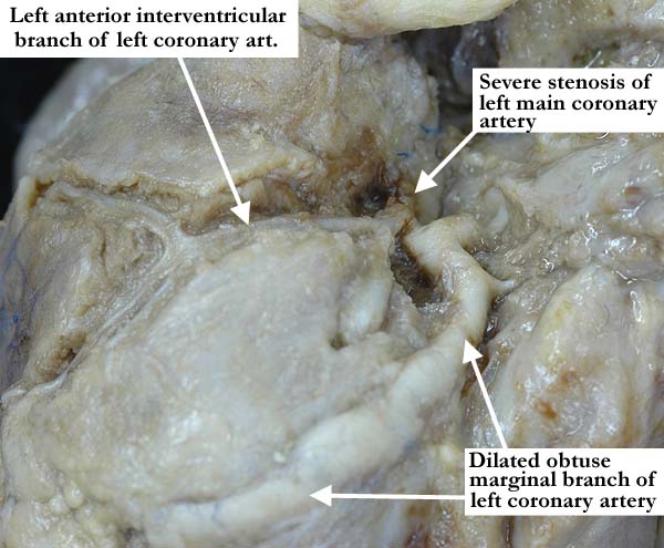
Modality: Anatomic specimen Orientation: Surface view of the left ventricular coronary arterial system Description: A close up view of the image shown on the left panel reveals the severe stenosis of the left main coronary artery. The thickened, dilated obtuse marginal branch is very stenotic at its branch point from the left main coronary artery. Contributor: Diane Spicer, BS Institution: The Congenital Heart Institute of Florida (CHIF) Image Label: A091503-66b Image Certification: pending AWG Rating: pending |
|||
|
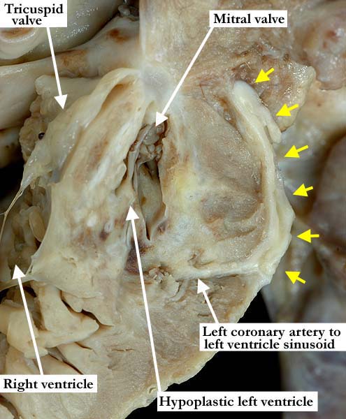
Modality: Anatomic specimen Orientation: Internal view of the left ventricle Description: This close up view simulates a four chamber echocardiographic view. There is endocardial fibroelastosis of the small left ventricle along with multiple, white fibrotic areas in the subendocardial region of both the ventricle and the interventricular septum. The thickened, dilated obtuse marginal branch (yellow arrows) of the left coronary artery has been bisected as it extends over the epicardial surface. Where it dives into the myocardium, a sinusoid is formed between it and the hypoplastic left ventricle. A thickened, markedly hypoplastic mitral valve guards the left ventricular inlet. Contributor: Diane Spicer, BS Institution: The Congenital Heart Institute of Florida (CHIF) Image Label: A091503-66c Image Certification: pending AWG Rating: pending
|
Modality: Angiocardiogram Orientation: Left ventricular injection (AP view) Description: This injection shows the hypoplastic left ventricle with opacification of the anterior interventricular coronary artery, filling from the left ventricle through ventricular-coronary fistulas. The hypoplastic aorta is filled by the abnormal connections in a retrograde fashion. Note the distance between the mass of the left ventricle and aorta. Contributor: Jorge M. Giroud, MD Institution: The Congenital Heart Institute of Florida (CHIF) Image Label: A091503-66d Source of Image: The Congenital Heart Institute of Florida (CHIF) Image Certification: pending AWG Rating: pending |
AWG Page Certification: pending
|
Copyright ipccc-awg.net All Rights Reserved. Frontpage-Templates.org |