|
||||||||
|
|
|||||||||
|
|||||||||
|
IPCCC Code: 01.01.09, 09.15.03, 06.02.92, [06.02.07], 07.06.04 |
|||
|
AEPC Derived Term: |
Hypoplastic left
heart syndrome (01.01.09) |
||
|
EACTS-STS Derived Term: |
Hypoplastic left heart syndrome (HLHS), Aortic atresia +
Mitral stenosis (01.01.09, 09.15.03, 06.02.92) |
||
|
ICD10 Derived Term: |
Hypoplastic left heart syndrome: Atresia, or marked
hypoplasia of aortic orifice or valve, with hypoplasia of ascending aorta
and defective development of left ventricle (with mitral valve stenosis or
atresia). (Q23.4) Endocardial fibroelastosis (I42.4) |
||
|
Comments: |
Please note that images A010109-30a, A010109-30b and A010109-30e are from different hearts. Images A010109-30c, A010109-30d are from the same heart. All hearts have varying degrees of left ventricular endocardial fibroelastosis. | ||
|
|
|||
|
Definition: NA |
|
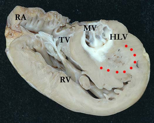
Modality: Anatomic specimen Orientation: Four chamber view viewed from the front Description: A four chamber view of this heart removed for transplantation shows a hypoplastic, fibroelastotic left ventricle (LV). The apical portion of this globular left ventricle demonstrates remnants of the trabecular component which are an unusual feature in HLHS (red dots). The mitral valve (MV) is severely stenotic, with short, fused tendinous cords. There is right ventricular hypertrophy and the right ventricle (RV) forms the apex of the heart. This image illustrates the endocardial fibroelastosis nicely. (TV-tricuspid valve, RA-right atrium). Contributor: Diane Spicer, BS Institution: The Congenital Heart Institute of Florida (CHIF) Image Label: A010109-30a Source of Image: Van Mierop Archive, University of Florida, Gainesville, FL Image Certification: 7 September 2013
AWG Rating:
|
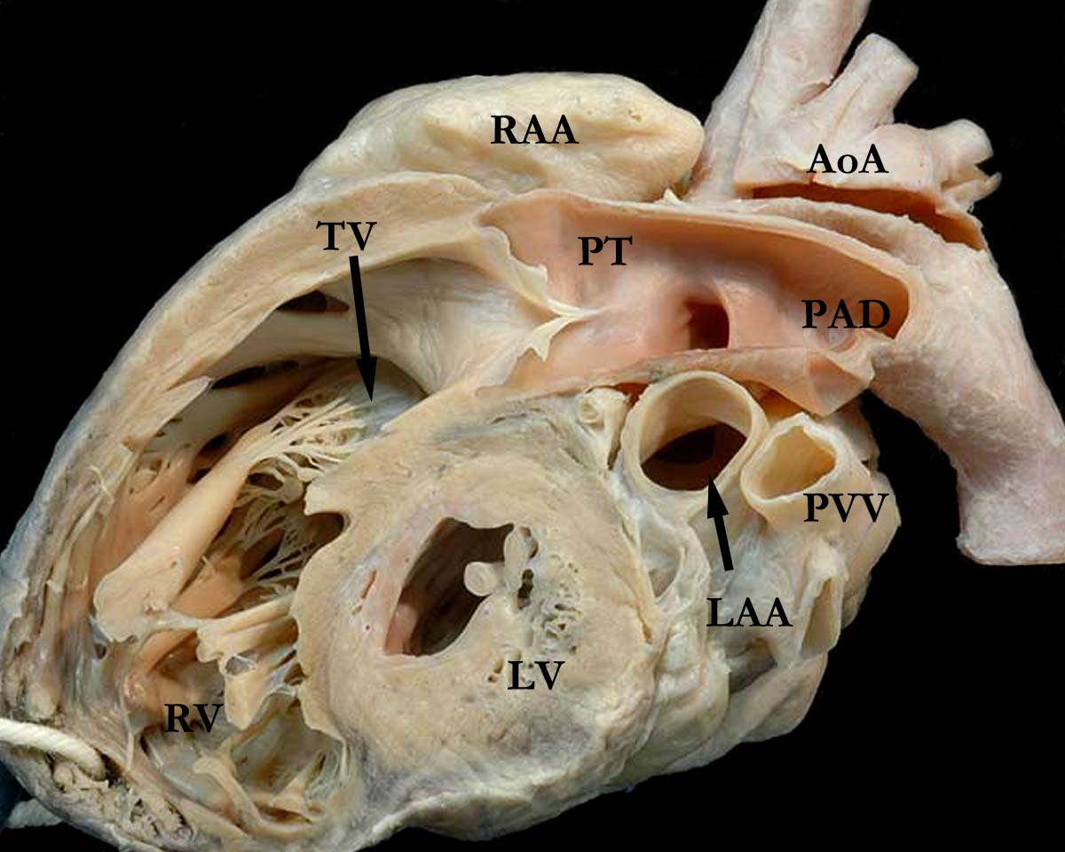
Modality: Anatomic specimen Orientation: Long axis view Description: A simulated right ventricular long axis echocardiographic view, cut from a different heart, illustrates a globular, hypoplastic left ventricle (LV). The right ventricle (RV) forms the apex of this heart, with the tricuspid valve (TV) in the inlet portion and a large pulmonary trunk (PT) forming the outlet component. The arterial duct is widely patent (PAD). The ascending aorta and aortic arch (AoA) are hypoplastic. Although not easily seen in this image, this left ventricle did have fibroelastotic changes. (RAA-right atrial appendage, LAA-left atrial appendage, PVV-pulmonary vein). Contributor: Diane Spicer, BS Institution: The Congenital Heart Institute of Florida (CHIF) Image Label: A010109-30b Source of Image: Van Mierop Archive, University of Florida, Gainesville, FL Image Certification: 7 September 2013
AWG Rating:
|
|||
|
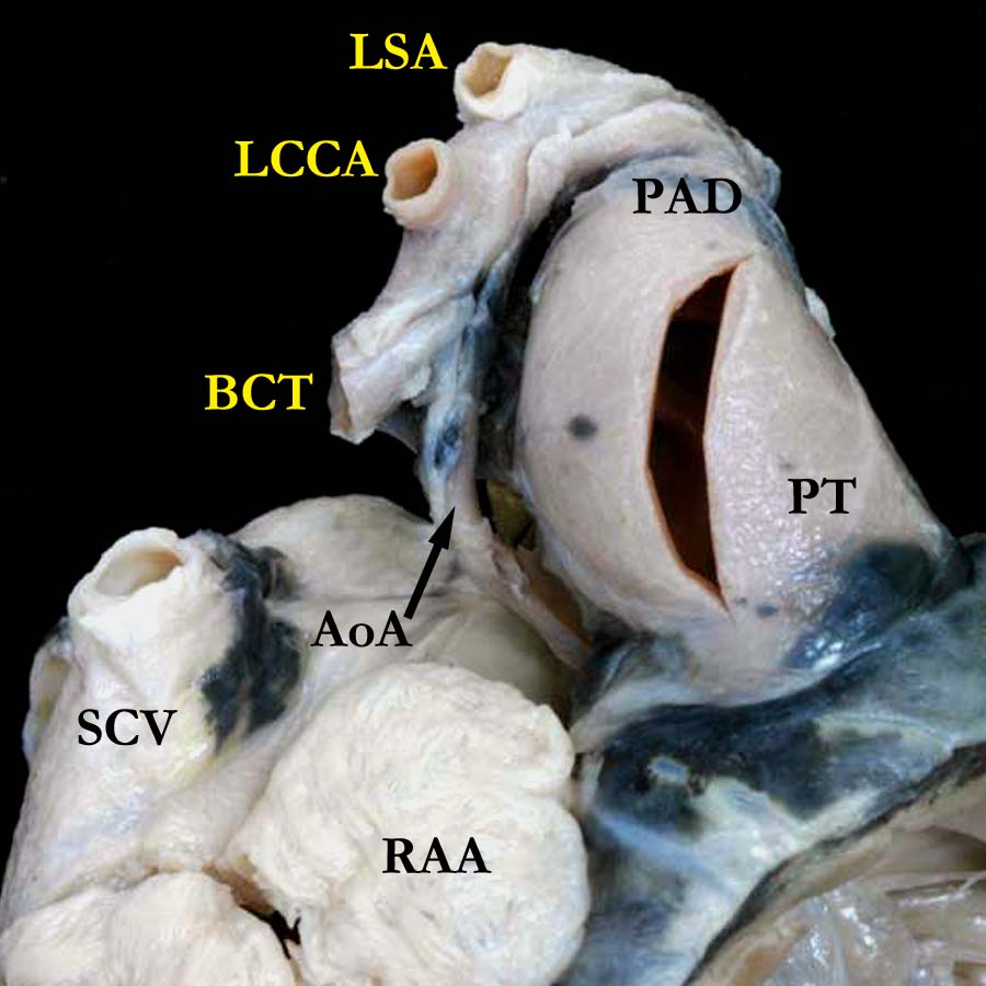
Modality: Anatomic specimen Orientation: Superior anterior view Description: The images of these two panels are taken from the same heart (refer to comments above). The anatomic view of the great vessels as they exit the base of the heart demonstrates a markedly hypoplastic ascending aorta (AoA) and a large, dilated pulmonary trunk (PT). The arterial duct (PAD) is widely patent. This is the classic appearance of the great vessels in a heart with hypoplastic left heart with aortic atresia and mitral atresia. In this particular case, the mitral valve had a pinpoint opening (please refer to image A010109-30d). The brachiocephalic trunk (BCT), the left carotid (LCCA) and the left subclavian (LSA) arteries exit the aortic arch in the usual fashion. (SCV-superior caval vein, RAA-right atrial appendage). Contributor: Diane Spicer, BS Institution: The Congenital Heart Institute of Florida (CHIF) Image Label: A010109-30c Source of Image: Van Mierop Archive, University of Florida, Gainesville, FL Image Certification: 7 September 2013
AWG Rating:
|
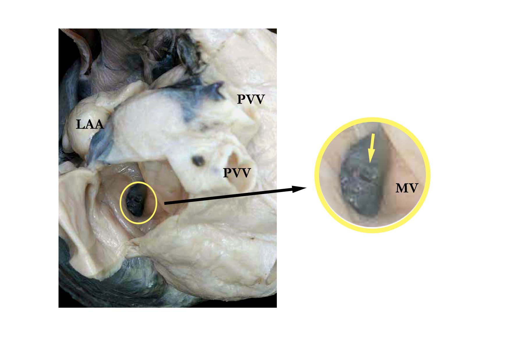
Modality: Anatomic specimen Orientation: Posterior left atrial view Description: This view of the mitral valve (MV) demonstrates severe stenosis of the left ventricular inlet to this heart with hypoplastic left ventricle. The pinpoint opening (yellow arrow) that can be seen rendered the valve functionally atretic. Note the dilated pulmonary veins (PVV). (LAA-left atrial appendage). Contributor: Diane Spicer, BS Institution: The Congenital Heart Institute of Florida (CHIF) Image Label: A010109-30d Source of Image: Van Mierop Archive, University of Florida, Gainesville, FL Image Certification: 7 September 2013
AWG Rating:
|
|||
|
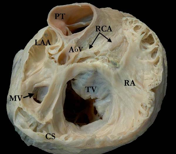
Modality: Anatomic specimen Orientation: Short axis view Description: This view of the base of a different heart with hypoplastic left heart, demonstrates the pertinent anatomic features of aortic valve (AoV) atresia and mitral valve (MV) stenosis. The right coronary artery (RCA) easily visualized extending to the right atrioventricular groove. The pulmonary trunk (PT) is large and dilated. The tricuspid valve (TV) guards the right ventricular inlet and the right atrium (RA) is dilated. There is severe stenosis of the mitral valve (MV) and the left atrium is hypoplastic. (LAA-left atrial appendage). Contributor: Diane Spicer, BS Institution: The Congenital Heart Institute of Florida (CHIF) Image Label: A010109-30e Source of Image: Van Mierop Archive, University of Florida, Gainesville, FL Image Certification: 7 September 2013
AWG Rating:
|
|
|||
AWG Certification: 7 September 2013
|
Copyright ipccc-awg.net All Rights Reserved. Frontpage-Templates.org |