|
||||||||
|
|
|||||||||
|
|||||||||
|
IPCCC: 06.06.01, 06.05.64, 06.05.25 |
|||
|
AEPC Derived Term: |
AVSD: isolated atrial component (primum ASD)(partial) (06.06.01) Left AV valvar hypoplasia in AVSD (06.05.64) Double orifice of left AV valve in AVSD (06.05.25) |
||
|
EACTS-STS Derived Term: |
ASD, Primum (06.06.01) AVC (AVSD)-modifier, AVSD AV valvar abnormality, L AV valvar hypoplasia in AVSD (06.05.64) AVC (AVSD)-modifier, AVSD AV valvar leaflet abnormality, Double orifice of L AV valve in AVSD (06.05.25) |
||
|
ICD10 Term: |
Atrioventricular septal defect (Q21.2) | ||
|
Definition: A congenital cardiac malformation that is a variant of an atrioventricular septal defect (atrioventricular canal) with an interatrial communication just above the atrioventricular valve, no interventricular communication just below the atrioventricular valve, separate right and left atrioventricular valvar orifices, and varying degrees of malformation of the left sided component of the common atrioventricular valve. The bridging leaflets of the common atrioventricular valve are bound down to the crest of the scooped out ventricular septum so that the potential for shunting through the atrioventricular septal defect is possible only at the atrial level and not at the ventricular level.
|
|
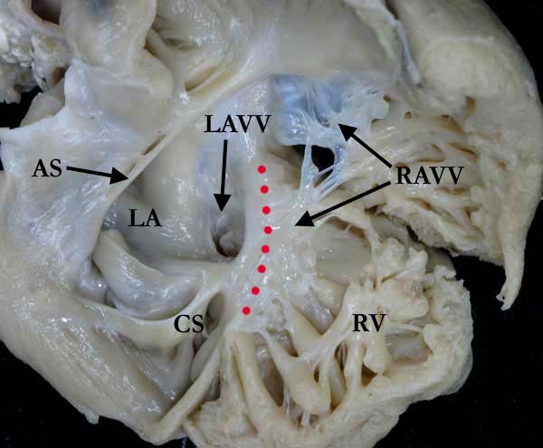
Modality: Anatomic specimen Orientation: Right ventricular view Description: The right ventricular (RV) free wall has been reflected, showing the larger right (RAVV) component of the common atrioventricular valve. This defect is unbalanced at the valvar level, the left side of the common valve appearing hypoplastic from the right side. The bridging leaflets are attached across the crest of the ventricular septum (red dots). The atrial septum (AS) is deficient, the atrial component of this defect quite large. (CS-coronary sinus, LA-left atrium, LV-left ventricle) Contributor: Diane Spicer, BS Institution: The Congenital Heart Institute of Florida (CHIF) Image Label: A060601-14a Source of Image: Van Mierop Archive, University of Florida, Gainesville, Florida Image Certification: 5 August 2011
AWG Rating:
|
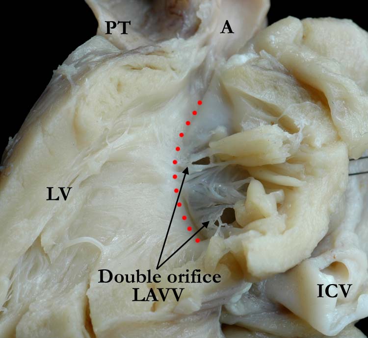
Modality: Anatomic specimen Orientation: Left ventricular long axis Description: The close up view of 3b clearly demonstrates the double orifice of the left component (LAVV) of the common atrioventricular valve, supported by two separate papillary muscles. The common atrioventricular valve is adherent along the entire length of the ventricular septum (red dots). There is fibrous continuity between the common atrioventricular valve and the aortic (A) valve and the subaortic outflow is very narrow. (ICV-inferior caval vein, LV-left ventricle, PT-pulmonary trunk) Contributor: Diane Spicer, BS Institution: The Congenital Heart Institute of Florida (CHIF) Image Label: A060601-14b Source of Image: Van Mierop Archive, University of Florida, Gainesville, Florida Image Certification: 5 August 2011
AWG Rating:
|
|||
AWG Page Certification: 5 August 2011
|
Copyright ipccc-awg.net All Rights Reserved. Frontpage-Templates.org |