|
|||||
|
|
||||||
|
||||||
|
IPCCC: 01.01.03, 02.01.02, 09.30.26, 06.06.00, 04.01.01 [04.01.02], 04.04.02, 04.05.01, 12.31.03 or 01.01.03, 09.05.11, 02.01.02, 09.30.26, 06.06.00, 04.01.02,
04.04.02, 04.01.03, 04.05.01 |
|||
|
AEPC Derived Term: |
Congenitally corrected transposition of great arteries (discordant AV & VA connections) (01.01.03) Dextrocardia: heart predominantly in right hemithorax (02.01.02) Right aortic arch (mirror image) branching pattern (09.30.26) Atrioventricular septal defect (06.06.00) Left superior caval vein (SVC) persisting to coronary sinus (04.01.01) vs Left superior caval vein (SVC) persisting to left-sided atrium (04.01.02) Coronary sinus defect in left atrium: completely unroofed (04.04.02) Absent bridging (innominate) vein (04.05.01) Modified right Blalock interposition shunt, (12.31.03) |
||
|
EACTS-STS Derived Term: |
Pulmonary atresia-VSD-modifier for intracardiac anatomy, AV Discordance, Aorta from RV, VA discordance: Corrected TGA with Pulmonary atresia (01.01.03, 09.05.11) Dextrocardia (right sided ventricular mass) (heart predominantly in right hemithorax) (02.01.02) Aortic arch, Right aortic arch (mirror image) branching pattern (09.30.26) AVC (AVSD) (06.06.00) Systemic venous anomaly, SVC, Bilateral SVC, LSVC to left-sided atrium (completely unroofed CS), Innominate absent, (04.01.02, 04.04.02, 04.01.03, 04.05.01) Palliation, Shunt - systemic-to-pulmonary, Blalock-Taussig (BTS), Modified Blalock-Taussig (MBTS) (deLeval shunt) (GOS shunt), Right (MBTSR) |
||
|
Definition: pending
|
|
Modality: Anatomic specimen Orientation: Anterior view Description: The apex of the heart lies to the right in this anterior anatomic view. The morphologically right atium is right-sided and the morphologically left atrium is left-sided, with bilateral superior caval veins. The left brachiocephalic vein is absent. The aortic arch extends to the right, with a left-sided arterial duct (yellow arrow). There is a modified Blalock-Taussig shunt (red dots) connecting the base of the right subclavian artery to the right main pulmonary artery (red asterisk). The tiny pulmonary trunk (yellow dots) lies just posterior to the aorta. Contributor: Diane E. Spicer, BS Institution: The Congenital Heart Institute of Florida (CHIF) Image Label: A010103-91a Image Source: Idriss Archive, Childrens Memorial Hospital, Chicago, IL Image Certification: pending AWG Rating: pending
|
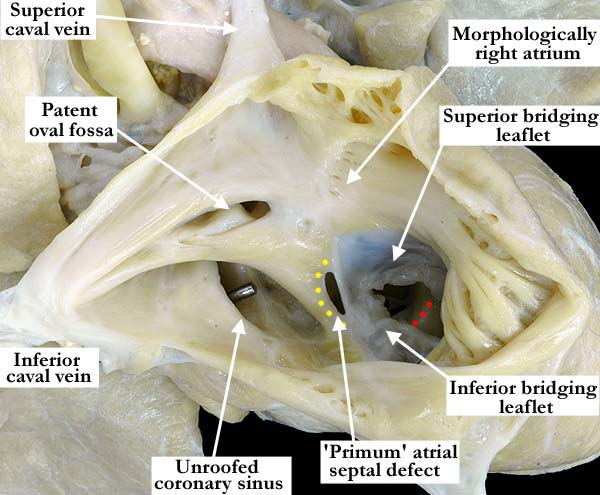
Modality: Anatomic specimen Orientation: Lateral view Description: The lateral view of this right-sided, morphologically right atrium reveals pectinate muscles extending to the mouth of the large coronary sinus. The coronary sinus is unroofed and the probe demonstrates an interatrial communication. The upper portion of the atrial septum is well formed with patent oval fossa. There is a common atrioventricular valve with the superior and inferior bridging leaflets attached leaving well defined atrial and ventricular components at mid defect. The so-called primum atrial septal defect is well defined with yellow dots marking the superior border of the defect. The scooped out portion of the ventricular septum is marked with red dots. Contributor: Diane E. Spicer, BS Institution: The Congenital Heart Institute of Florida (CHIF) Image Label: A010103-91b Image Source: Idriss Archive, Childrens Memorial Hospital, Chicago, IL Image Certification: pending AWG Rating: pending
|
|||
|
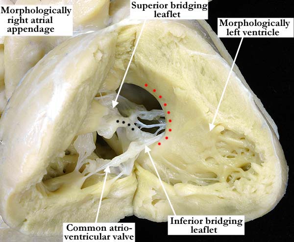
Modality: Anatomic specimen Orientation: Posterior view Description: The morphologically left ventricle has been opened in a clam-shell like fashion and lies posterior to the morphologically right ventricle. There are discordant atrioventricular connections with the right-sided, morphologically right atrium connected to the morphologically left ventricle, the common atrioventricular valve in the inlet portion. The ventricular component of the atrioventricular septal defect shows the characteristic scooped-out appearance (red dots) of the ventricular septum. The black dots on the common atrioventricular valve illustrate the area of fusion between the superior and inferior bridging leaflets. Contributor: Diane E. Spicer, BS Institution: The Congenital Heart Institute of Florida (CHIF) Image Label: A010103-91c Image Source: Idriss Archive, Childrens Memorial Hospital, Chicago, IL Image Certification: pending AWG Rating: pending
|
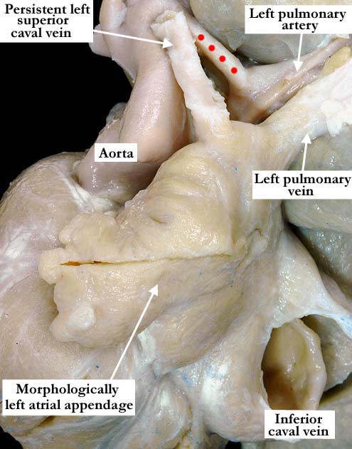
Modality: Anatomic specimen Orientation: Posterior lateral view Description: This posterior lateral view of the left-sided, morphologically left atrium and atrial appendage shows the persistent left superior caval vein entering the roof of the atrium. The pulmonary venous drainage is normal. The left-sided, arterial duct (red dots) connects the aorta to the left pulmonary artery. Contributor: Diane E. Spicer, BS Institution: The Congenital Heart Institute of Florida (CHIF) Image Label: A010103-91d Image Source: Idriss Archive, Childrens Memorial Hospital, Chicago, IL Image Certification: pending AWG Rating: pending
|
|||
|
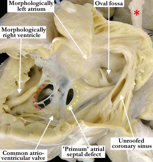
Modality: Anatomic specimen Orientation: Left lateral view Description: This left, posterior lateral view of the opened left-sided, morphologically left atrium demonstrates the normal appearance of the oval fossa, along with the unroofed coronary sinus, forming an interatrial communication. The persistent left superior caval vein is marked with a red asterisk. There are discordant atrioventricular connections, the left-sided, morphologically left atrium connected to a morphologically right ventricle. There is a common atrioventricular valve in the inlet. The superior and inferior bridging leaflets are fused, this creating well defined atrial and ventricular components of the defect. The atrial border of the so-called primum portion of the defect is outlined with yellow dots. The red dots mark the scooped-out portion of the ventricular septum. Contributor: Diane E. Spicer, BS Institution: The Congenital Heart Institute of Florida (CHIF) Image Label: A010103-91e Image Source: Idriss Archive, Childrens Memorial Hospital, Chicago, IL Image Certification: pending AWG Rating: pending
|
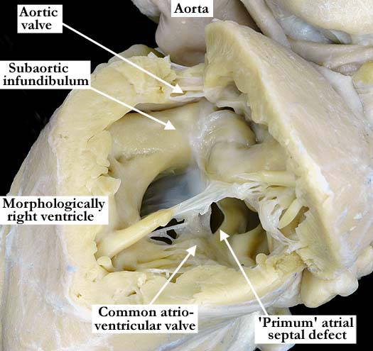
Modality: Anatomic specimen Orientation: Anterior view Description: The anterior-most ventricle is a morphologically right ventricle and has been opened in a clam-shell like fashion to demonstrate the outlet component. Looking from the apex, the common atrioventricular valve lies within the inlet and is separated from the aortic valve by the subaortic, muscular infundibulum. The right ventricle and the infundibulum are hypertrophied. There is a discordant ventriculo-arterial connection, the aorta connected to the morphologically right ventricle. Contributor: Diane E. Spicer, BS Institution: The Congenital Heart Institute of Florida (CHIF) Image Label: A010103-91f Image Source: Idriss Archive, Childrens Memorial Hospital, Chicago, IL Image Certification: pending AWG Rating: pending |
|||
AWG Page Certification: pending
|
Copyright ipccc-awg.net All Rights Reserved. Frontpage-Templates.org |