|
|||||
|
|
||||||
|
||||||
|
IPCCC: 07.15.21, 07.11.02, 07.10.04 |
|||
|
AEPC Derived Term: |
Multiple VSDs: 2 (07.15.21) Muscular VSD in inlet septum (07.11.02) Perimembranous VSD with extension to right ventricular outlet (cranial) (07.10.04) |
||
|
EACTS-STS Derived Term: |
VSD, Multiple, 2 VSD (07.15.21) VSD, Type 4 (Muscular), Inlet (Posterior) (07.11.02) VSD, Type 2 (Perimembranous) (Paramembranous) (Conoventricular), Outlet (07.10.04) |
||
|
|
Definitions:
Commentary:
Common Synonyms: |
|
|
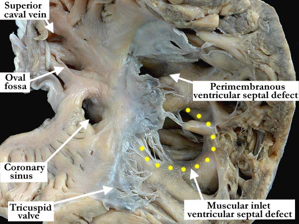
Modality: Anatomic specimen Orientation: Right ventricle Description: This anterior anatomic view of the right atrium and ventricle demonstrates two ventricular septal defects. The larger defect is a muscular inlet defect (yellow dots) that extends onto the mid interventricular septum and is quite large. The defect has complete muscular borders and is clearly separated from the perimembranous ventricular septal defect by a large muscle bar. At the upper border of the perimembranous ventricular septal defect there is a cleft in the tricuspid valve. The oval fossa is small and the coronary sinus is dilated secondary to a persistent left superior caval vein which is not shown in this illustration Contributor: Diane Spicer, BS Institution: The Congenital Heart Institute of Florida (CHIF) Image Label: A071521-70a Source of Image: Image Certification: pending AWG Rating: pending
|
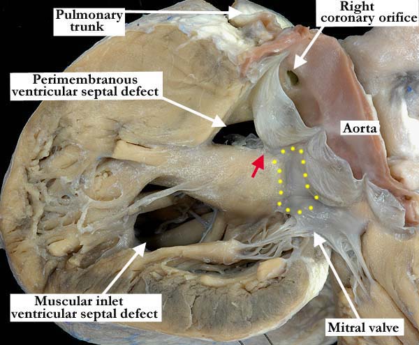
Modality: Anatomic specimen Orientation: Left ventricle Description: The left ventricular view of the heart shown in the left panel clearly demonstrates the borders of the two ventricular septal defects. The inlet defect is surrounded by muscle and is separated from the perimembranous defect by a large, septal bar of muscle. The perimembranous defect lies beneath the right coronary leaflet of the aortic valve and borders a small portion of the membranous septum (red arrow). Note that the majority of the membranous septum (yellow dots) is intact. Contributor: Diane Spicer, BS Institution: The Congenital Heart Institute of Florida (CHIF) Image Label: A071521-70b Source of Image: Image Certification: pending AWG Rating: pending
|
|||
AWG Page Certification: pending
|
Copyright ipccc-awg.net All Rights Reserved. Frontpage-Templates.org |