|
|||||
|
|
||||||
|
||||||
IPCCC Code: 07.11.04
AEPC Code: Muscular VSD in central trabecular septum
EACTS-STS Code: VSD, Type 4 (Muscular), Trabecular, Midventricular (Midmuscular)
|
|
|
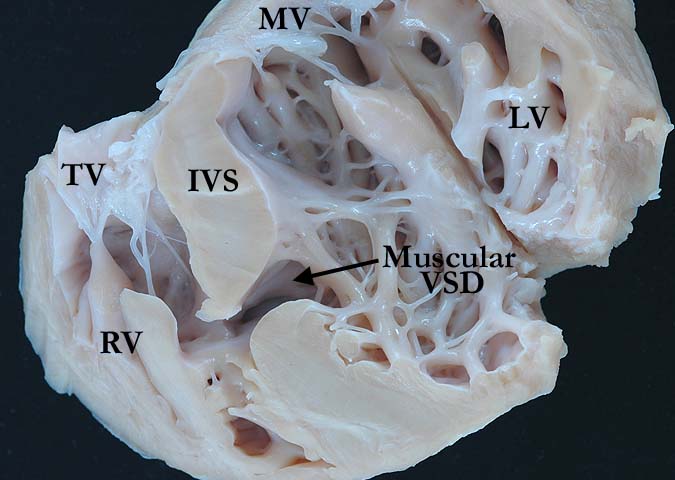
This four chamber view demonstrates a muscular ventricular septal defect (VSD) at mid interventricular septum (IVS). The borders of the defect are entirely muscular. (TV-tricuspid valve, RV-right ventricle, MV-mitral valve, LV-left ventricle)
Contributor: Diane Spicer, BS Image Name: ACHS97-3943bcopy.jpg |
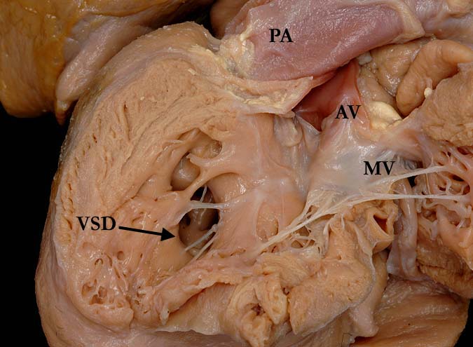
Left ventricular (LV) view of a mid septal muscular ventricular septal defect (VSD). This defect would not be amendable to device closure with the trabeculations traversing the opening. (MV-mitral valve, AV-aortic valve, PA-pulmonary artery)
Contributor: Diane Spicer, BS Image Name: UF65-63bcopy.jpg
|
|||
|
|
||||
|
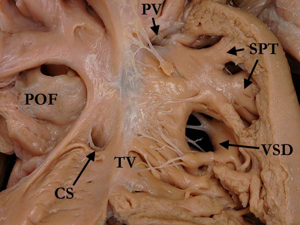
This view of the right ventricular (RV) septum shows a mid septal ventricular septal defect (VSD). The borders are entirely muscular. There are prominent septoparietal trabeculations (SPT) that mimic ventricular septal defect, with no interventricular communication (OF-oval fossa, TV-tricuspid valve, PV-pulmonary valve)
Contributor: Diane Spicer, BS Image Name: UF65-63acopy.jpg
|
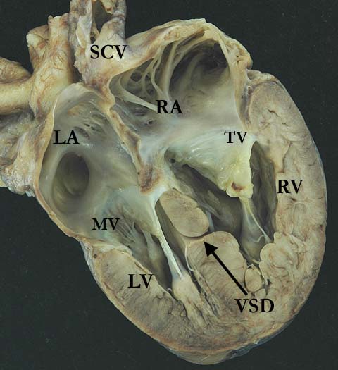
The four chamber view of this heart demonstrates a mid septal, restrictive, muscular septal defect (VSD). The interatrial septum is intact and there is biventricular hypertrophy. (SCV-superior caval vein, RA-right atrium, LA-left atrium, RV-right ventricle, LV-left ventricle)
Contributor: Diane Spicer, BS Image Name: MVSDrestr.jpg
|
|||
AWG Certification: Pending
|
Copyright ipccc-awg.net All Rights Reserved. Frontpage-Templates.org |