|
|||||
|
|
||||||
|
||||||
|
IPCCC: 07.11.02 |
|||
|
AEPC Derived Term: |
Muscular VSD in inlet septum (07.11.02) | ||
|
EACTS-STS Derived Term: |
VSD, Type 4 (Muscular), Inlet (Posterior) (07.11.02) | ||
|
Definition: A muscular ventricular septal defect located beneath the septal leaflet of the tricuspid valve.
Commentary: Some refer to this ventricular septal defect as a
posterior muscular ventricular septal defect. |
|
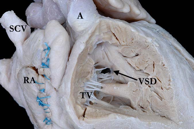
Modality: Anatomic specimen Orientation: Right ventricle Description: This view of the right ventricle demonstrates the anatomic features of transposition of the great arteries with the tricuspid valve (TV) in the inlet and the aorta (A) forming the outlet portion. There is an inlet, muscular ventricular septal defect (VSD). Multiple sutures extend across the right atrium (RA), this heart status post Mustard procedure. (SCV-superior caval vein). Additional diagnosis = IPCCC: 01.05.01. AEPC: Discordant VA connections (TGA). EACTS-STS: TGA (Transposition of the Great Arteries) (Concordant atrioventricular connections and Discordant ventriculo-arterial connections) Contributor: Diane Spicer, BS Institution: The Congenital Heart Institute of Florida (CHIF) Image Label: A071102-44a Source of Image: Van Mierop Archive, University of Florida, Gainesville, Florida Image Certification: pending AWG Rating: pending
|
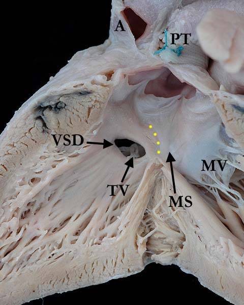
Modality: Anatomic specimen Orientation: Left ventricle Description: The left ventricular view of the heart seen in the left panel, shows the pulmonary trunk within the outlet of this morphologically left ventricle in this heart with transposition of the great vessels. A pulmonary artery band is present. There is a muscular inlet ventricular septal defect (VSD), the border of the intact membranous septum (MS) demonstrated with the yellow dots. A portion of the tricuspid valve (TV) is seen through the ventricular septal defect. The fibrous continuity between the pulmonary valve and the mitral valve (MV) is easily appreciated. (A-aorta) Contributor: Diane Spicer, BS Institution: The Congenital Heart Institute of Florida (CHIF) Image Label: A071102-44b Source of Image: Van Mierop Archive, University of Florida, Gainesville, Florida Image Certification: pending AWG Rating: pending
|
|||
|
|
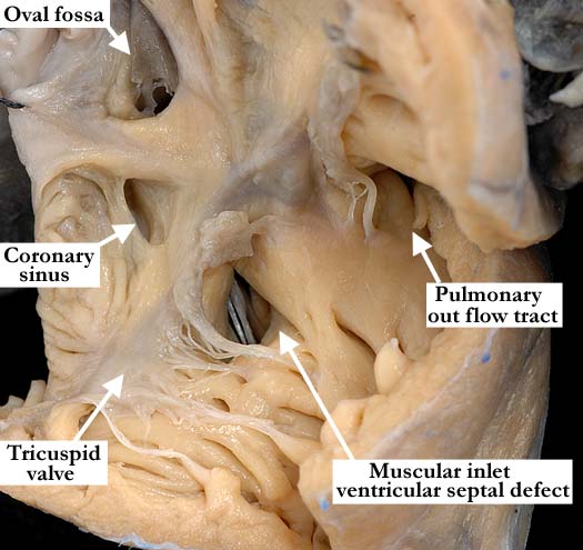
Modality: Anatomic specimen Orientation: Anterior right ventricle Description: This anterior anatomic view demonstrates a muscular inlet ventricular septal defect beneath the septal leaflet of the tricuspid valve. The defect is entirely surrounded by muscle and lies within the inlet portion of the interventricular septum. There are concordant atrioventricular connections with a small defect in the flap valve of the oval fossa. Contributor: Diane Spicer, BS Institution: The Congenital Heart Institute of Florida (CHIF) Image Label: A071102-44c Source of Image: Image Certification: pending AWG Rating: pending
|
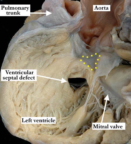
Modality: Anatomic specimen Orientation: Left ventricle Description: The left ventricular view of the muscular inlet ventricular septal defect seen from the right side in the image from the left panel clearly demonstrates the muscular borders of the defect and the intact membranous septum (yellow dots). Contributor: Diane Spicer, BS Institution: The Congenital Heart Institute of Florida (CHIF) Image Label: A071102-44d Source of Image: Image Certification: pending AWG Rating: pending |
||
|
|
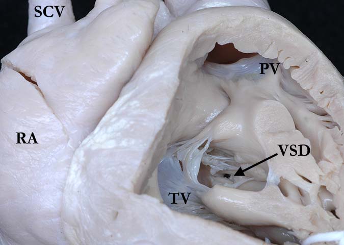
Modality: Anatomic specimen Orientation: Anterior right ventricle Description: This close up, anatomical view of the right ventricle shows the intact inlet portion, guarded by the tricuspid valve (TV) with a small, restrictive, inlet, muscular ventricular septal defect (VSD). (SCV-superior caval vein, RA-right atrium, PV-pulmonary valve) Contributor: Diane Spicer, BS Institution: The Congenital Heart Institute of Florida (CHIF) Image Label: A071102-44e Source of Image: Van Mierop Archive, University of Florida, Gainesville, Florida Image Certification: pending AWG Rating: pending
|
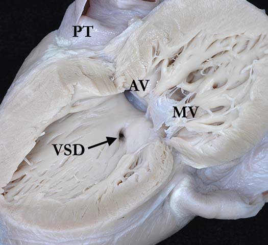
Modality: Anatomic specimen Orientation: Left ventricle Description: The left ventricle of the same heart shown in the left pane is opened in clam shell fashion, demonstrating the mitral valve (MV) in its inlet, the aortic valve (AV) within the outlet and a small, restrictive, muscular ventricular septal defect (VSD) Contributor: Diane Spicer, BS Institution: The Congenital Heart Institute of Florida (CHIF) Image Label: A071102-44f Source of Image: Van Mierop Archive, University of Florida, Gainesville, Florida Image Certification: pending AWG Rating: pending |
||
|
|
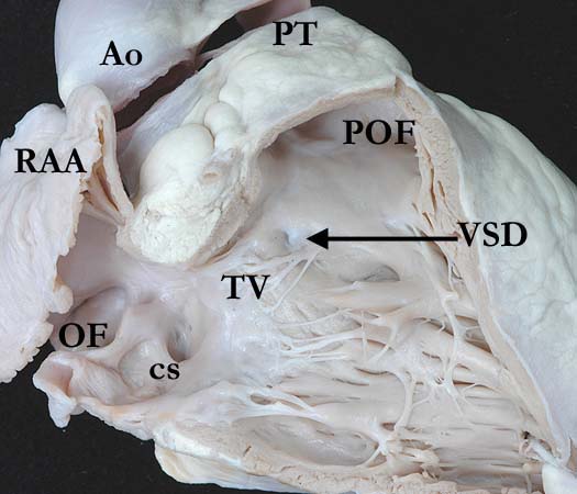
Modality: Anatomic specimen Orientation: Anterior right ventricle Description: This anatomic view of the septal surface of this morphologically right ventricle shows the tricuspid valve (TV) in the inlet and the pulmonary trunk (PT) extending from the pulmonary outflow tract (POF). Just superior to the cordal attachments that extend from the medial papillary muscle to the septal leaflet of the tricuspid valve, there is a tiny, probe patent, but most likely functionally closed muscular ventricular septal defect (VSD). (RAA-right atrial appendage, OF-oval fossa, cs-coronary sinus, Ao-aorta). Additional diagnosis = IPCCC: 07.16.03. AEPC: Spontaneous closure of VSD by fibromuscular reaction. EACTS-STS: VSD-modifier, Spontaneous closure of VSD, By fibromuscular reaction. Contributor: Diane Spicer, BS Institution: The Congenital Heart Institute of Florida (CHIF) Image Label: A071102-44g Source of Image: Van Mierop Archive, University of Florida, Gainesville, Florida Image Certification: pending AWG Rating: pending
|
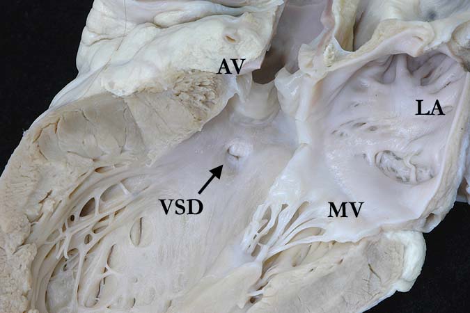
Modality: Anatomic specimen Orientation: Left ventricle Description: A long axis view of the left atrium (LA) and left ventricle of the heart shown in the left panel shows the mitral valve (MV) in the inlet portion and the aortic valve (AV) in the outlet. Just below the right coronary leaflet of the aortic valve there is a tiny, probe patent, most likely functionally closed, muscular ventricular septal defect (VSD) Contributor: Diane Spicer, BS Institution: The Congenital Heart Institute of Florida (CHIF) Image Label: A071102-44h Source of Image: Van Mierop Archive, University of Florida, Gainesville, Florida Image Certification: pending AWG Rating: pending
|
||
AWG Page Certification: pending
|
Copyright ipccc-awg.net All Rights Reserved. Frontpage-Templates.org |