|
|||||
|
|
||||||
|
||||||
|
IPCCC: 07.10.08 [+/- 07.10.01, 07.17.05], 12.14.02, 12.24.23 |
|||
|
AEPC Derived Term: |
VSD + malaligned outlet septum anteriorly: overriding subaortic outflow (Fallot type) (07.10.08) Pulmonary trunk band (PA band) (12.14.02) Ligation of patent arterial duct (PDA)(12.24.23) |
||
|
EACTS-STS Derived Term: |
VSD, Type 2 (Perimembranous) (Paramembranous) (Conoventricular), Outlet, Conal septal malalignment, TOF type (07.10.01, 07.17.05) VSD-modifier, VSD + malaligned outlet septum anteriorly: overriding aortic valve (Fallot type) (07.10.08) PA banding (12.14.02) PDA closure, Surgical therapy, Ligation (12.24.23) |
||
|
Definition: A VSD with the muscular outlet (infundibular)
septum, or its fibrous remnant, deviated antero-superiorly in the presence
of overriding of the orifice of the subaortic outflow
Commentary: This VSD is usually found with concordant ventriculo-arterial
connections or alignments, as in tetralogy of Fallot |
|
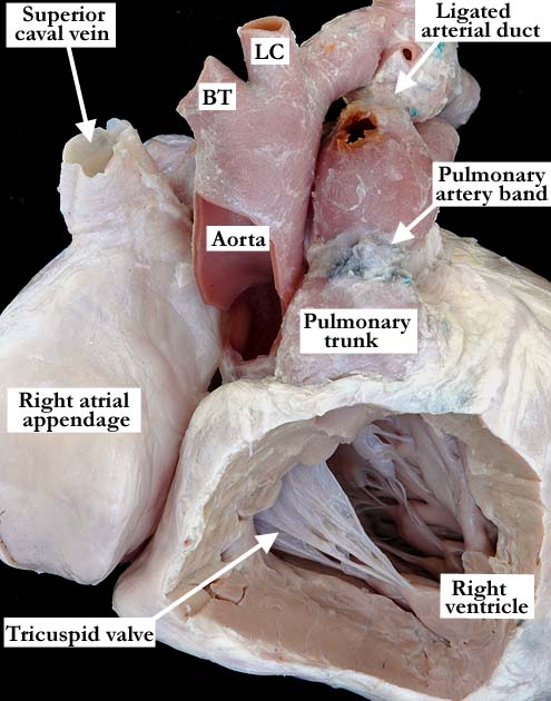
Modality: Anatomic specimen Orientation: Anterior superior view Description: This view of the great vessels as they exit the ventricular mass shows the aorta arising slightly to the right of its usual position. The pulmonary artery is dilated with a pulmonary artery band in place and a ligated arterial duct. The right atrium is dilated and there is right ventricular hypertrophy. Contributor: Diane Spicer, BS Institution: The Congenital Heart Institute of Florida (CHIF) Image Label: A071008-74a Source of Image: Van Mierop Archive, University of Florida, Gainesville, FL Image Certification: pending AWG Rating: pending
|
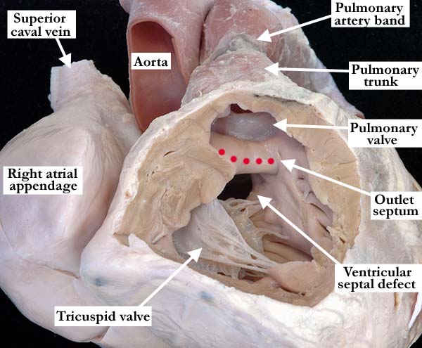
Modality: Anatomic specimen Orientation: Right ventricular view Description: The anterior apical view of the right ventricle demonstrates malalignment of the outlet septum into the right ventricle with an unobstructed subpulmonary outlet. The aorta overrides the ventricular septum, although the aortic valve is not appreciated in this view. This defect is consistent with an Eisenmenger type of ventricular septal defect. Contributor: Diane Spicer, BS Institution: The Congenital Heart Institute of Florida (CHIF) Image Label: A071008-74b Source of Image: Van Mierop Archive, University of Florida, Gainesville, FL Image Certification: pending AWG Rating: pending
|
|||
|
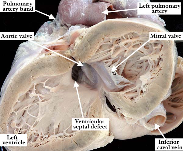
Modality: Anatomic specimen Orientation: Left ventricular view Description: In this anatomic view of the left ventricle, the free wall has been lifted away to demonstrate the aortic root. The aorta clearly overrides the interventricular septum with a good portion of the aortic valve supported in the right ventricle. Contributor: Diane Spicer, BS Institution: The Congenital Heart Institute of Florida (CHIF) Image Label: A071008-74c Source of Image: Van Mierop Archive, University of Florida, Gainesville, FL Image Certification: pending AWG Rating: pending
|
|
|||
AWG Page Certification: pending
|
Copyright ipccc-awg.net All Rights Reserved. Frontpage-Templates.org |