|
|||||
|
|
||||||
|
||||||
IPCCC Code: 07.10.03
AEPC Code: Perimembranous VSD with extension to right ventricular
trabecular component (anterior)
EACTS-STS Code: VSD, Type 2 (Perimembranous) (Paramembranous) (Conoventricular), Trabecular
|
|
|
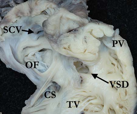
This is a right ventricular view of a perimembranous ventricular septal defect that extends into the trabecular component with clefting of the septal leaflet of the tricuspid valve (TV). The medial papillary muscule arises from the apex of the defect with very short, fused cordal attachments in this area. This defect has a small component that extends into the inlet component. The free wall of the morphologically right ventricle has been removed and the right atrium is open showing a normal coronary sinus (CS) and superior caval vein (SCV), along with a patent oval foramen. The pulmonary valve (PV) is unobstructed.
Contributor: Diane Spicer, BS Image Name: UF01-07acopy.jpg |
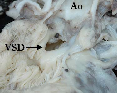
The left ventricular view of this perimembranous ventricular septal defect shows the characteristic features of a defect that extends into the trabecular component, with the cleft in the tricuspid valve easily seen through the defect. A few blood cysts are present at the superior rim of the defect and adjacent to the right coronary leaflet.
Contributor: Diane Spicer, BS Image Name: UF01-07bcopy.jpg
|
|||
|
|
||||
|
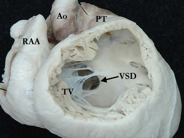
Perimembranous VSD with extension to the inlet and trabecular components. The cordal attachments of the tricuspid valve (TV) are seen around the anterior superior rim of the defect. (RAA-right atrial appendage, PT-pulmonary trunk, Ao-aorta)
Contributor: Diane Spicer, BS Image Name: UFHM413bcopy.jpg |
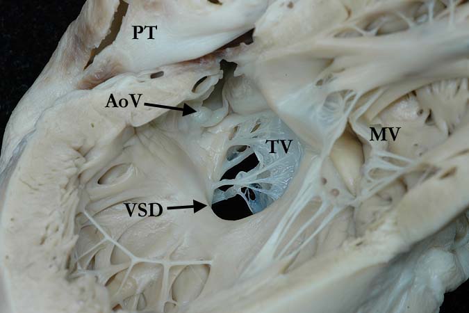
This left ventricular view of a perimembranous ventricular septal defect extends to the inlet and trabecular components. The aortic (AoV), tricuspid valve (TV) and mitral valve (MV) are all in fibrous continuity. The cordal attachments of the TV can easily be seen along the edge of the defect. The non-coronary leaflet of the aortic valve appears distant from the defect which compares with the anatomy seen in perimembranous ventricular septal defects that extend to the inlet component.
Contributor: Diane Spicer, BS Image Name: UFHM413ecopy.jpg
|
|||
AWG Certification: Pending
|
Copyright ipccc-awg.net All Rights Reserved. Frontpage-Templates.org |