|
|||||
|
|
||||||
|
||||||
|
IPCCC: 09.01.03, 07.10.14, 09.27.10 |
|||
|
AEPC Derived Term: |
Common arterial trunk (truncus) + separate origin of pulmonary arteries (type II) (09.01.03) VSD + overriding truncal valve (absent outlet septum) with inferior muscular rim (07.10.14) Left arterial ligament (09.27.10) |
||
|
EACTS-STS Derived Term: |
Truncus arteriosus, With both PA's coming from truncus and unobstructed arch, Collett and Edwards II - separate origin of PA's, (09.01.03), Truncus arteriosus-modifier, Truncus arteriosus with VSD, With inferior muscular rim (not extending to membranous septum) (07.10.14) Ligamentum arteriosum-modifier for sidedness, Left ligamentum arteriosum (09.27.10) |
||
|
Definition: pending
|
|
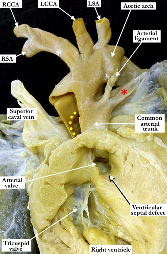
Modality: Anatomic specimen Orientation: Anterior view Description: The right ventricular free wall has been windowed. In this anterior inferior view from the apex, the right ventricular inlet and outlet components are clearly visualized. The tricuspid valve lies in the inlet. There is a subarterial ventricular septal defect and a common arterial trunk valve guarding the outlet. The ventricular septal defect is restrictive and has a posterior inferior muscular rim. Arising from the right aspect of the common arterial trunk is the aortic component. There is a normal left aortic arch with the usual branching pattern of the brachiocephalic arteries. The right (yellow dots) and left (red asterisk) pulmonary arteries have a separate, although closely approximated, origin from the posterior aspect of the common arterial trunk. There is an arterial ligament arising at the point where the left main pulmonary artery bifurcates from the common trunk. Contributor: Diane E. Spicer, BS Institution: The Congenital Heart Institute of Florida (CHIF) Image Label: A090103-90a Image Source: Medical University of South Carolina Image Certification: pending AWG Rating: pending
|
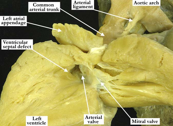
Modality: Anatomic specimen Orientation: Left ventricular view Description: The left ventricular view demonstrates the restrictive, subarterial ventricular septal defect. The common arterial trunk valve overrides the ventricular septum and is in fibrous continuity with the mitral valve. There is left ventricular hypertrophy. Contributor: Diane E. Spicer, BS Institution: The Congenital Heart Institute of Florida (CHIF) Image Label: A090103-90b Image Source: Medical University of South Carolina Image Certification: pending AWG Rating: pending |
|||
AWG Page Certification: pending
|
Copyright ipccc-awg.net All Rights Reserved. Frontpage-Templates.org |