|
|||||
|
|
||||||
|
||||||
|
IPCCC
Code:
06.01.34, 07.05.06,
06.01.51 |
|||
|
AEPC Code: |
Ebstein's malformation of tricuspid valve, Right ventricular outflow tract obstruction due to AV valve, Tricuspid papillary muscle abnormality (for Ebst2b) |
||
|
EACTS-STS Code: |
Tricuspid valve disease, Ebstein's anomaly, RVOT abnormality, RVOTO, RV outflow tract obstruction due to AV valve, Tricuspid valve disease, Tricuspid valve pathology, Tricuspid papillary muscle abnormality (for Ebst2b) |
||
|
Definition: NA |
|
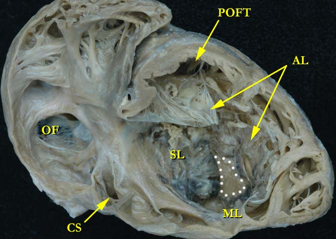
In this view, the anterior wall of the right atrium and right ventricle have been removed to show the severe Ebstein malformation of the tricuspid valve. The white dots illustrate the only remaining portion of the right ventricular septal surface that has not been covered by the tricuspid valve. The inferior aspect of the right ventricular myocardium has been atrialized. The anterior leaflet extends over the pulmonary outlet, with only a small opening allowing blood flow into the pulmonary artery, effectively causing severe pulmonary stenosis. (from the University of Florida Van Mierop Archive)
Contributor: Diane Spicer, BS Image Name: Ebst1a.jpg
|
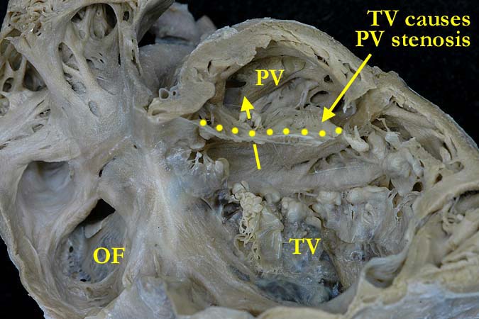
A close up view of Ebst1a showing the anterior leaflet of the tricuspid valve extending over the pulmonary outlet portion of the right ventricle. A yellow arrow demonstrates the tiny hole that remains in this leaflet. (from the University of Florida Van Mierop Archive)
Contributor: Diane Spicer, BS Image Name: Ebst1b.jpg |
|||
|
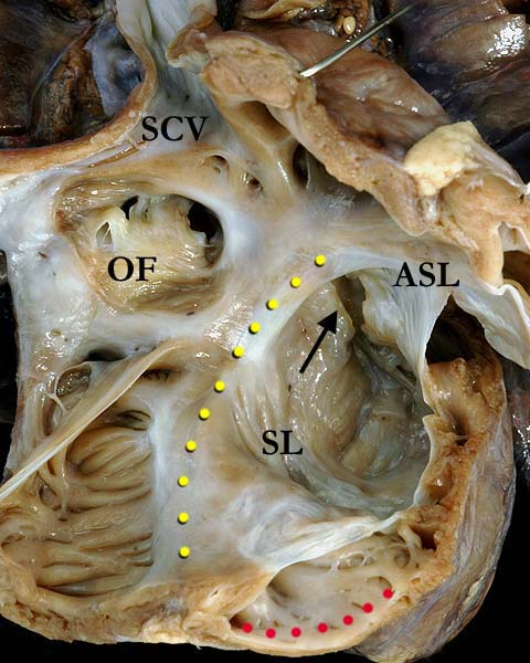
This anatomical view of the right ventricle shows the rotational displacement of the septal (SL) and mural leaflets of the tricuspid valve from the inner curve of the heart. The yellow dots illustrate where the tricuspid valve annulus should be. The red dots outline the atrialized portion of the right ventricle. The majority of the septal and mural leaflets of the tricuspid valve are adherent to the septal surface of the right ventricle. The anterior superior leaflet (ASL) has been partially cut and is sail-like. The black arrow illustrates the keyhole outlet to the pulmonary outflow. (SCV-superior caval vein, OF-oval fossa)
Contributor: Diane Spicer, BS Image Name: Ebst2a.jpg
|
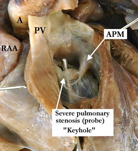
The pulmonary outflow of the heart shown in Ebst2a illustrating the keyhole outlet to the pulmonary trunk. The probe extends through this opening, the tricuspid valve slightly thickened in this area. There is an aberrant papillary muscle (APM) supporting this portion of the tricuspid valve.
Contributor: Diane Spicer, BS Image Name: Ebst2b.jpg |
|||
AWG Certification: Pending
|
Copyright ipccc-awg.net All Rights Reserved. Frontpage-Templates.org |