|
||||||||
|
|
|||||||||
|
|||||||||
|
IPCCC: 06.01.03, 06.01.49, 05.01.22, 05.04.02 |
|||
|
AEPC Derived Term: |
Tricuspid valvar dysplasia (06.01.03) Tricuspid valve primary chords absent (06.01.49) Eustachian valve prolapsing through tricuspid valve (05.01.22) Atrial septal defect (ASD) within oval fossa (secundum) (05.04.02) |
||
|
EACTS-STS Derived Term: |
Tricuspid valve disease, Tricuspid valve pathology, Mucoid thickening (Tricuspid valve dysplasia) (06.01.03) Tricuspid valve disease, Tricuspid valve pathology, Tricuspid chordal abnormality, Tricuspid valve primary chords absent (06.01.49) Atrial abnormality, RA abnormality, Eustachian valve prolapsing through tricuspid valve (05.01.22) ASD, Secundum (05.04.02) |
||
|
Definition: pending
|
|
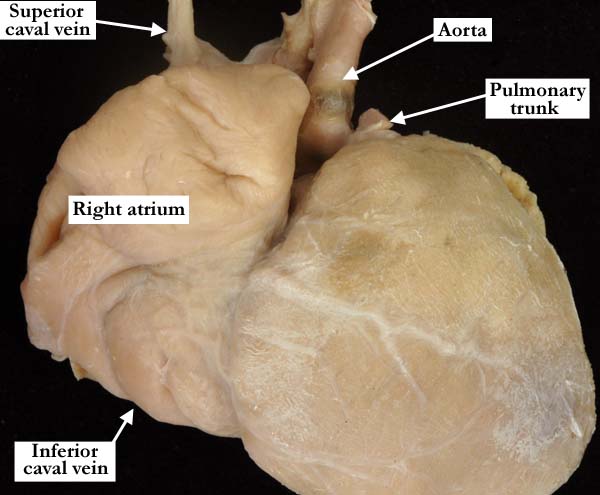
Modality: Anatomic specimen Orientation: Anterior view Description: The right atrium and atrial appendage are markedly dilated. Contributor: Diane E. Spicer, BS Institution: The Congenital Heart Institute of Florida (CHIF) Image Label: A060103-96a Source of Image: Van Mierop Archive, University of Florida Image Certification: pending AWG Rating: pending
|
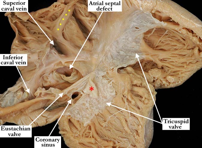
Modality: Anatomic specimen Orientation: Anterior view Description: The free wall of the right ventricle has been folded back demonstrating the right atrial dilatation and the dysplastic tricuspid valve. The tricuspid valve is inserted directly onto the septal surface and to the tips of the papillary muscles. The only visible tendinous cords are those attaching to the medial papillary muscle. The Eustachian valve is large and redundant with a windsock-like structure (red asterisk) that prolapses through the tricuspid valve. There is a secundum, atrial septal defect. Contributor: Diane E. Spicer, BS Institution: The Congenital Heart Institute of Florida (CHIF) Image Label: A060103-96b Source of Image: Van Mierop Archive, University of Florida Image Certification: pending AWG Rating: pending
|
|||
|
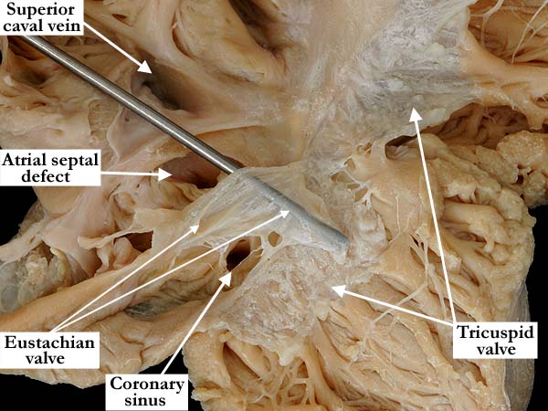
Modality: Anatomic specimen Orientation: Close up, anterior view Description: A close up view of the dysplastic tricuspid valve and the portion of the Eustachian valve that prolapses through the valve. There is a secundum, atrial septal defect. Contributor: Diane E. Spicer, BS Institution: The Congenital Heart Institute of Florida (CHIF) Image Label: A060103-96c Source of Image: Van Mierop Archive, University of Florida Image Certification: pending AWG Rating: pending
|
||||
AWG Page Certification: pending
|
Copyright ipccc-awg.net All Rights Reserved. Frontpage-Templates.org |