|
||||||||
|
|
|||||||||
|
|||||||||
|
IPCCC: 01.01.02, 12.01.41, 12.14.02 |
|||
|
AEPC Derived Term: |
Transposition of great arteries (concordant AV & discordant VA connections) & IVS (01.01.02) Balloon atrial septostomy by pull back (Rashkind) (12.01.41) Pulmonary trunk band (PA band) (12.14.02) |
||
|
EACTS-STS Derived Term: |
TGA - IVS (Transposition of the Great Arteries - Intact Ventricular Septum) (Concordant atrioventricular connections and Discordant ventriculo-arterial connections - IVS) (01.01.02) Cardiovascular catheterization procedure, Therapeutic, Perforation (establishing interchamber and/or intervessel communication), Atrial septum PA banding (12.14.02) |
||
|
ICD 10 Term: |
Discordant ventriculoarterial connection (Q20.3) | ||
|
Definition: pending
Comments: This series of images illustrate the anatomic and diagnostic features of Transposition of the Great Arteries with Intact Ventricular Septum. Using specimens from different collections, various therapeutic maneuvers are demonstrated with their corresponding IPCCC terms. |
|
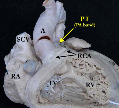
Modality: Anatomic specimen Orientation: Anterior view Description: This view shows the aorta (A) anterior to the pulmonary trunk (PT) in this heart with transposition of the great arteries. There is a pulmonary artery band in place. There are concordant atrioventricular connections, the morphologically right atrium (RA) connected to the morphologically right ventricle (RV) in the usual fashion with the tricuspid valve (TV) guarding the inlet. The ventriculoarterial connections are discordant, the aorta within the outlet component of the right ventricle. The right coronary artery (RCA) has an aberrant origin and extends anterior to the aorta to the right atrioventricular groove. Contributor: Diane Spicer, BS Institution: The Congenital Heart Institute of Florida (CHIF) Image Label: A010102-55a Source of Image: Van Mierop Archive, University of Florida, Gainesville, Florida Image Certification: pending AWG Rating: pending
|
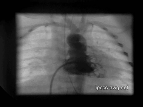
Modality: Angiocardiogram Orientation: Anterior-posterior view Description: Right ventricular angiogram of a newborn with transposition of the great arteries. The aorta arises from the right ventricle. Contributor: Otto N. Krogmann, MD Institution: Paedetric Cardiology - Heart Center Duisburg, Germany Image Label: An010102-55b Source of Image: Paedetric Cardiology - Heart Center Duisburg, Germany Image Certification: pending AWG Rating: pending
|
|||
|
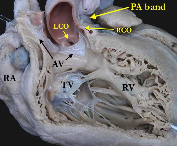
Modality: Anatomic specimen Orientation: Anterior view Description: A close up view of the heart shown in the left panel with the anterior portion of the aortic valve (AV) opened to demonstrate the discordant atrioventricular connections consistent with transposition of the great arteries. The aorta is anterior to the pulmonary trunk with a pulmonary artery (PA) band in place. The right coronary orifice (RCO) is adjacent to a commissure. Note the muscular infundibulum separating the aortic valve from the tricuspid valve (TV). (RA-right atrium). Contributor: Diane Spicer, BS Institution: The Congenital Heart Institute of Florida (CHIF) Image Label: A010102-55c Source of Image: Van Mierop Archive, University of Florida, Gainesville, Florida Image Certification: pending AWG Rating: pending
|
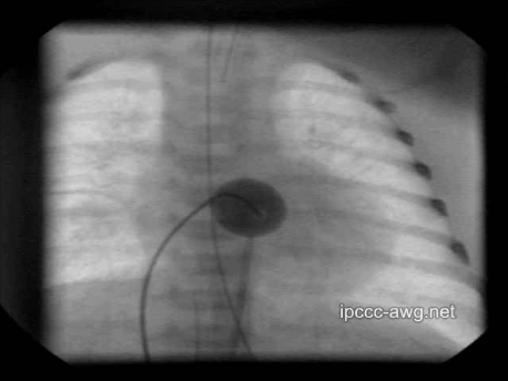
Modality: Fluoroscopy of balloon septostomy Orientation: Anterior-posterior view Description: In a newborn with transposition of the great arteries a Rashkind maneuver is performed to increase the interatrial communication. A Rashkind balloon catheter is inflated with 1 to 3 ml of a 50% dye-saline mixture in the left atrium and rapidly withdrawn into the right atrium hereby tearing the interatrial septum. Contributor: Otto N. Krogmann, MD Institution: Paedetric Cardiology - Heart Center Duisburg, Germany Image Label: An010102-55d Source of Image: Paedetric Cardiology - Heart Center Duisburg, Germany Image Certification: pending AWG Rating: pending |
|||
|
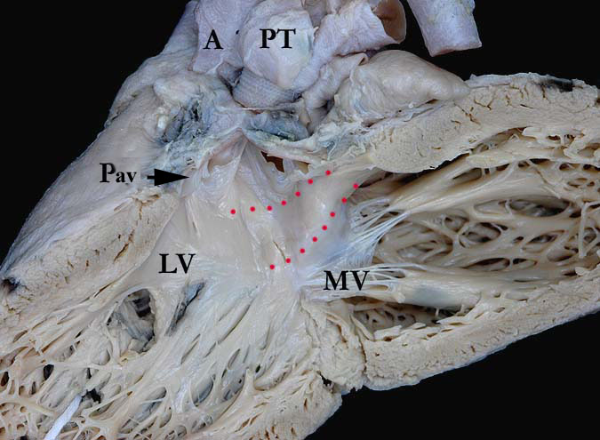
Modality: Anatomic specimen Orientation: Left ventricular view Description: The left ventricle of the heart shown above demonstrates the concordant atrioventricular connections with the mitral valve (MV) guarding the inlet to the morphologically left ventricle (LV) and the discordant ventriculoarterial connections with the pulmonary trunk (PT) forming the outlet portion of the morphologically left ventricle. Note the muscular infundibulum (red dots) separating the mitral valve from the pulmonary valve (Pav). Within the ventricular septum, there is an artifactual defect. Contributor: Diane Spicer, BS Institution: The Congenital Heart Institute of Florida (CHIF) Image Label: A010102-55c Source of Image: Van Mierop Archive, University of Florida, Gainesville, Florida Image Certification: pending AWG Rating: pending
|
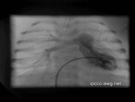
Modality: Angiocardiogram Orientation: Anterior-posterior view Description: Left ventricular angiogram of a newborn with transposition of the great arteries. The pulmonary artery arises from the left ventricle. Contributor: Otto N. Krogmann, MD Institution: Paedetric Cardiology - Heart Center Duisburg, Germany Image Label: An010102-55f Source of Image: Paedetric Cardiology - Heart Center Duisburg, Germany Image Certification: pending AWG Rating: pending |
|||
AWG Page Certification: pending
|
Copyright ipccc-awg.net All Rights Reserved. Frontpage-Templates.org |