|
||||||||
|
|
|||||||||
|
|||||||||
|
IPCCC Code: 01.01.01 |
|||
|
AEPC Code: |
Tetralogy of Fallot | ||
|
EACTS-STS Code: |
Tetralogy of Fallot | ||
|
Definition: NA |
|
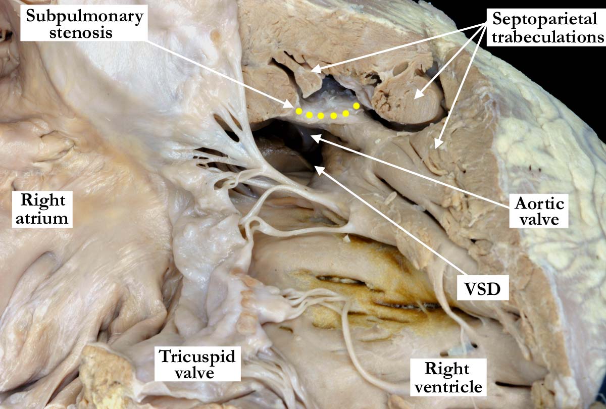
Modality: Anatomic specimen Orientation: Anterior view of the right ventricle Description: This close up, anatomical view of this morphologically right ventricle demonstrates the classic features of tetralogy of Fallot. The thickened tricuspid valve guards the inlet to the right ventricle which is hypertrophic. There is a perimembranous ventricular septal defect (VSD) and because the aorta overrides the interventricular septum, the aortic valve can be seen at the roof of the ventricular septal defect. Anterior deviation of the outlet septum (yellow dots) causes the infundibular or subpulmonary stenosis. Hypertrophy of the septoparietal trabeculations contributes to the subpulmonary stenosis. Contributor: Diane Spicer, BS Institution: The Congenital Heart Institute of Florida (CHIF) Image Label: A010101-36a Source of Image: Van Mierop Archive, University of Florida, Gainesville, FL Image Certification: pending AWG Rating:
|
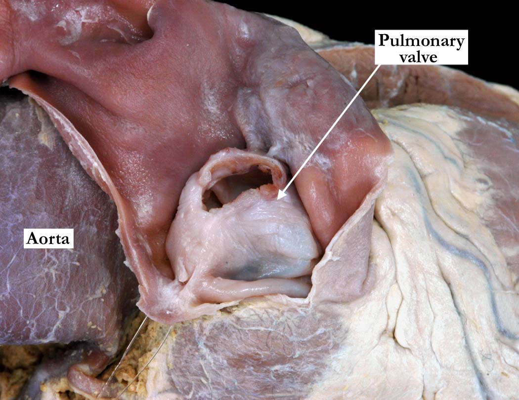
Modality: Anatomic specimen Orientation: Superior view of the pulmonary valve Description: This close up view of the pulmonary valve shows a thickened, stenotic, dome-shaped pulmonary valve. Contributor: Diane Spicer, BS Institution: The Congenital Heart Institute of Florida (CHIF) Image Label: A010101-36b Source of Image: Van Mierop Archive, University of Florida, Gainesville, FL Image Certification: pending AWG Rating: |
|||
|
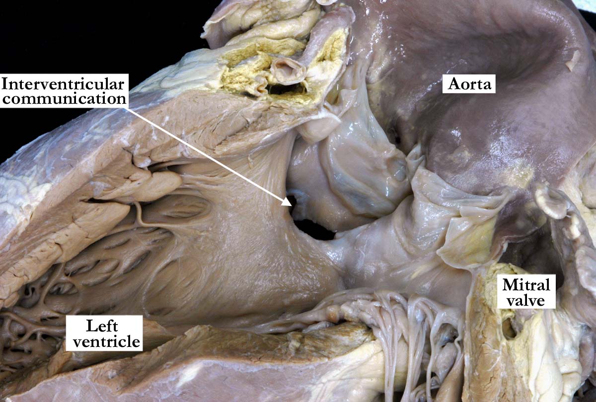
Modality: Anatomic specimen Orientation: Lateral view of the left ventricle Description: The left ventricular aspect of the heart shown above demonstrates the interventricular communication and the aorta overriding the interventricular septum. The aortic valve is in fibrous continuity with the mitral and tricuspid valves (not shown). Contributor: Diane Spicer, BS Institution: The Congenital Heart Institute of Florida (CHIF) Image Label: A010101-36c Source of Image: Van Mierop Archive, University of Florida, Gainesville, FL Image Certification: pending AWG Rating:
|
|
|||
AWG Certification: Pending
|
Copyright ipccc-awg.net All Rights Reserved. Frontpage-Templates.org |