|
||||||||
|
|
|||||||||
|
|||||||||
|
IPCCC: 04.06.00, 04.06.03, Q1.01.51, 04.08.11 |
|||
|
AEPC Derived Term: |
Totally anomalous
pulmonary venous connection: supracardiac (04.06.00) Obstructed pulmonary venous connection due to extrinsic compression (04.08.11) |
||
|
EACTS-STS Derived Term: |
Total anomalous
pulmonary venous connection (TAPVC), Type 1 (supracardiac) (04.06.00) Pulmonary venous connection obstructed-modifier for type of obstruction, Obstructed pulmonary venous connection, Extrinsic compression (04.08.11) |
||
|
ICD 10 Term: |
Total anomalous pulmonary venous connection (Q26.2) | ||
|
Definition: pending
|
|
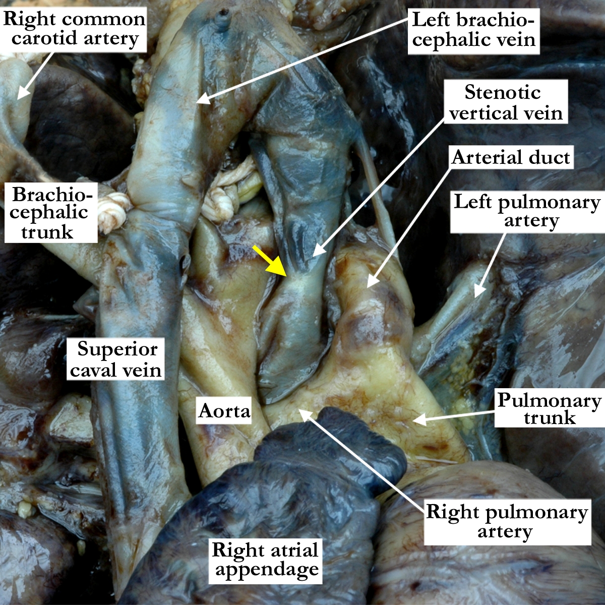
Modality: Anatomic specimen Orientation: Anterior view Description: This close up anterior view demonstrates supracardiac totally anomalous pulmonary venous connection. The pulmonary veins form a confluence posterior to the heart (see image A040600-125b) that continues superiorly as a vertical vein, which is narrowed (yellow arrow) as it runs between the aorta and pulmonary trunk before draining into the left brachiocephalic vein. Contributor: Diane E. Spicer, BS Institution: The Congenital Heart Institute of Florida (CHIF) Image Label: A040600-125a Source of Image: The Congenital Heart Institute of Florida (CHIF) Image Certification: pending AWG Rating: pending
|
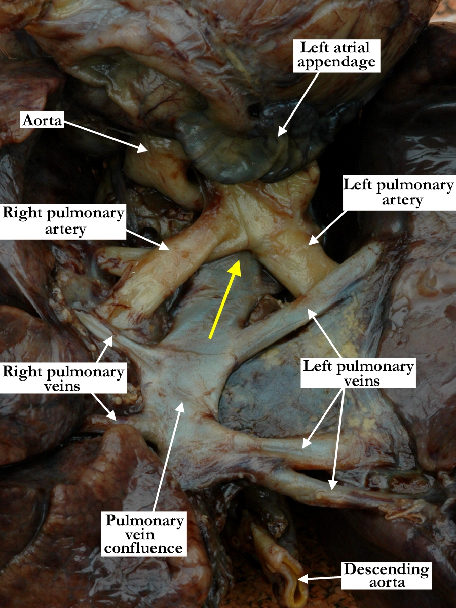
Modality: Anatomic specimen Orientation: Posterior inferior view Description: The heart is lifted away from the lungs to show the right and left pulmonary veins forming a venous confluence that gives rise to a vertical vein (yellow arrow) that extends superiorly between the right and left pulmonary arteries and between the aorta and pulmonary trunk, where it becomes stenotic (see image A040600-125a). Note the dilated pulmonary venous confluence proximal to the area of obstruction in the pulmonary venous pathway. Contributor: Diane E. Spicer, BS Institution: The Congenital Heart Institute of Florida (CHIF) Image Label: A040600-125b Source of Image: The Congenital Heart Institute of Florida (CHIF) Image Certification: pending AWG Rating: pending |
|||
AWG Page Certification: pending
|
Copyright ipccc-awg.net All Rights Reserved. Frontpage-Templates.org |