|
||||||||
|
|
|||||||||
|
|||||||||
|
IPCCC: 04.06.00, 04.06.03, Q1.01.51 |
|||
|
AEPC Derived Term: |
Totally anomalous
pulmonary venous connection: supracardiac (04.06.00) |
||
|
EACTS-STS Derived Term: |
Total anomalous
pulmonary venous connection (TAPVC), Type 1 (supracardiac) (04.06.00) |
||
|
ICD 10 Term: |
Total anomalous pulmonary venous connection (Q26.2) | ||
|
Definition: pending
|
|
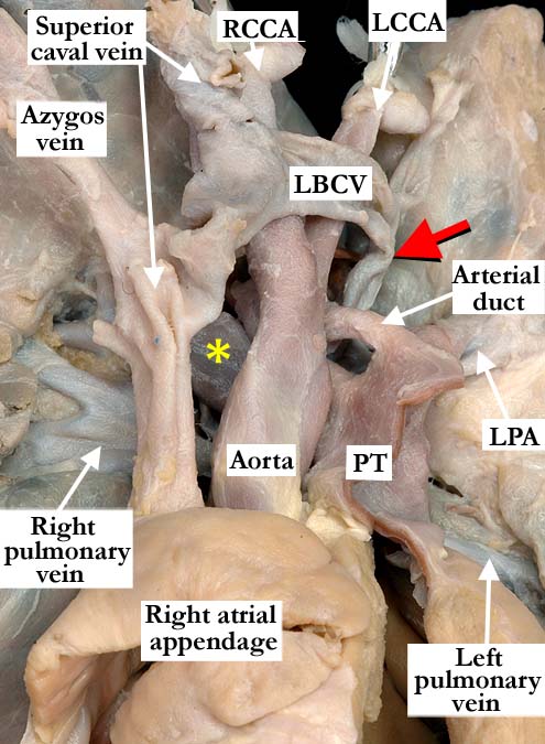
Modality: Anatomic specimen Orientation: Anterior view Description: The anterior anatomic view in this heart with concordant atrioventricular and ventriculo-arterial connections demonstrates a normally related aorta and pulmonary trunk (PT). The left brachiocephalic vein (LBCV) drains normally to the superior caval vein, and the azygos vein has been lifted from its usual position to extend the superior caval vein. There is supracardiac totally anomalous pulmonary venous return via a vertical vein (red arrow) to the left brachiocephalic vein. The right and left pulmonary veins join in a confluence (see image A040600-124b) posterior to the heart, the vertical vein then extending posterior to the arterial duct. (yellow asterisk- right pulmonary artery, LCCA-left common carotid artery, LPA-left pulmonary artery, RCCA-right common carotid artery) Contributor: Diane E. Spicer, BS Institution: The Congenital Heart Institute of Florida (CHIF) Image Label: A040600-124a Source of Image: Idriss Archive, Childrens Memorial Hospital, Chicago, IL Image Certification: pending AWG Rating: pending
|
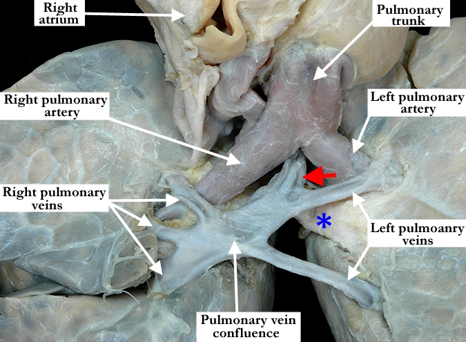
Modality: Anatomic specimen Orientation: Posterior inferior view Description: The heart has been lifted cephalad, using the so-called Taussig maneuver (reference below), to demonstrate the confluence between the right and left pulmonary veins, which is posterior to the heart. A vertical vein (red arrow) then extends from the confluence, running between the pulmonary arteries and the left bronchus, the so-called bronchopulmonary vice, before draining into the left brachiocephalic vein (see image A040600-124a). (Blue asterisk- left main bronchus) Contributor: Diane E. Spicer, BS Institution: The Congenital Heart Institute of Florida (CHIF) Image Label: A040600-124b Source of Image: Idriss Archive, Childrens Memorial Hospital, Chicago, IL Image Certification: pending AWG Rating: pending |
|||
|
In Pediatric Autopsy: Fetus, Newborn and Child (Ch 2). Editors: Enid Gilbert-Barness and Diane E. Debich-Spicer. Handbook of Pediatric Autopsy Pathology. 1st ed. Totowa, NJ: Humana Press;2005:17 |
||||
AWG Page Certification: pending
|
Copyright ipccc-awg.net All Rights Reserved. Frontpage-Templates.org |