|
||||||||
|
|
|||||||||
|
|||||||||
|
IPCCC: 4.08.30, 04.06.03, Q1.01.55, 04.06.12, Q1.01.52 |
|||
|
AEPC Derived Term: |
Totally anomalous
pulmonary venous connection: mixed (04.08.30) |
||
|
EACTS-STS Derived Term: |
Total anomalous
pulmonary venous connection (TAPVC), Type 4 (mixed) (04.08.30) |
||
|
ICD 10 Term: |
Total anomalous pulmonary venous connection (Q26.2) | ||
|
Definition: pending
|
|
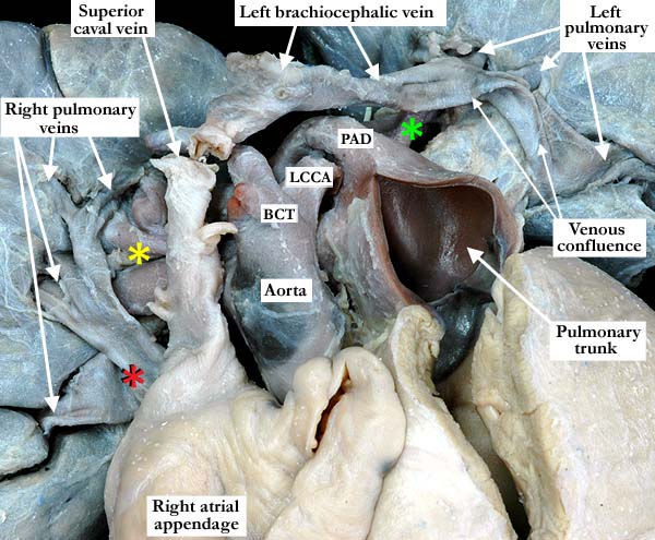
Modality: Anatomic specimen Orientation: Anterior view Description: The anterior anatomic view of this heart with concordant atrioventricular and ventriculo-arterial connections demonstrates normally related great arteries, and shows the left brachiocephalic vein draining to the superior caval vein. There is mixed totally anomalous pulmonary venous return, with the left pulmonary veins draining in supracardiac fashion to a venous confluence that then drains into the left brachiocephalic vein, while the right pulmonary veins (red asterisk) drain to the coronary sinus, and thence to the right atrium (see image A040830-123b) (yellow asterisk-right pulmonary artery, green asterisk-left pulmonary artery, BCT-brachiocephalic trunk, LCCA-left common carotid artery, PAD-patent arterial duct) Contributor: Diane E. Spicer, BS Institution: The Congenital Heart Institute of Florida (CHIF) Image Label: A040830-123a Source of Image: Idriss Archive, Childrens Memorial Hospital, Chicago, IL Image Certification: pending AWG Rating: pending
|
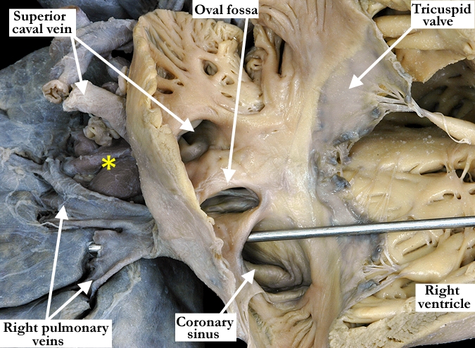
Modality: Anatomic specimen Orientation: Anterior lateral view Description: The right atrium has been opened, and a probe inserted through the coronary sinus to reveal the anomalous drainage of the venous return from the right lung. The veins from the left lung drain in supracardiac fashion to the left brachiocephalic vein. As expected, the orifice of the coronary sinus is markedly dilated. There are concordant atrioventricular connections, with the tricuspid valve guarding the inlet to the right ventricle. The oval foramen is probe patent. The course of the left pulmonary veins can be viewed in image A040830-123a. (yellow asterisk-right pulmonary artery) Contributor: Diane E. Spicer, BS Institution: The Congenital Heart Institute of Florida (CHIF) Image Label: A040830-123b Source of Image: Idriss Archive, Childrens Memorial Hospital, Chicago, IL Image Certification: pending AWG Rating: pending |
|||
AWG Page Certification: pending
|
Copyright ipccc-awg.net All Rights Reserved. Frontpage-Templates.org |