|
||||||||
|
|
|||||||||
|
|||||||||
|
IPCCC: 04.08.20, 04.06.22 |
|||
|
AEPC Derived Term: |
Totally anomalous
pulmonary venous connection: infracardiac (04.08.20) |
||
|
EACTS-STS Derived Term: |
Total anomalous
pulmonary venous connection (TAPVC), Type 3 (infracardiac) (04.08.20) |
||
|
ICD 10 Term: |
Total anomalous pulmonary venous connection (Q26.2) | ||
|
Definition: pending
|
|
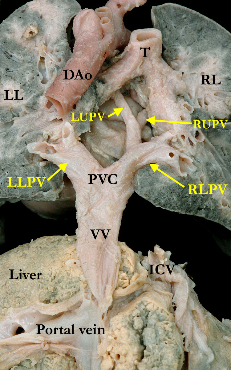
Modality: Anatomic specimen Orientation: Posterior view Description: This posterior view of the pulmonary veins illustrates total anomalous pulmonary venous connection below the diaphragm from the pulmonary vein confluence (PVC) via a descending vertical vein (VV) that drains to the portal vein. The right (RLPV) and left (LLPV) lower pulmonary veins are much larger than the upper pulmonary veins (RUPV, LUPV) as they enter the confluence. (DAo-descending aorta, T-trachea, RL-right lung, LL-left lung, ICV- inferior caval vein) Contributor: Diane E. Spicer, BS Institution: The Congenital Heart Institute of Florida (CHIF) Image Label: A040820-34a Source of Image: Van Mierop Archive, University of Florida, Gainesville, Florida Image Certification: 1 March 2014
AWG Rating:
|
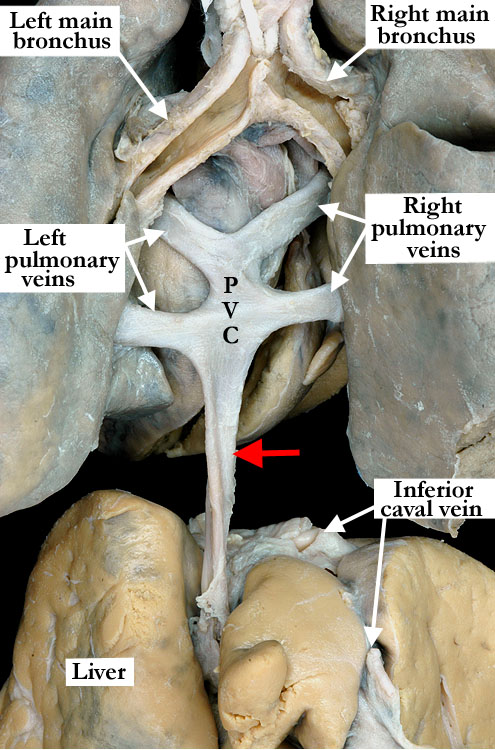
Modality: Anatomic specimen Orientation: Posterior view
Description: This posterior view shows
morphologically normal right and left main bronchuses and subdiaphragmatic
totally anomalous pulmonary venous connection. The right and left pulmonary
veins join a venous confluence (PVC)
that gives rise to a descending vertical vein (red arrow) and extends below
the diaphragm (previously removed) and although not imaged, connects to the
portal vein. Contributor: Diane E. Spicer, BS Institution: The Congenital Heart Institute of Florida (CHIF) Image Label:A040820-34b Source of Image: Idriss Archive, Ann & Robert H. Lurie Children's Hospital of Chicago, Chicago, IL Image Certification: 1 March 2014
AWG Rating:
|
|||
|
|
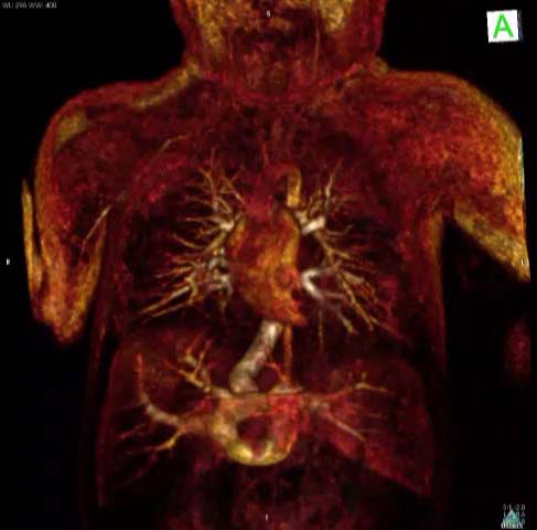
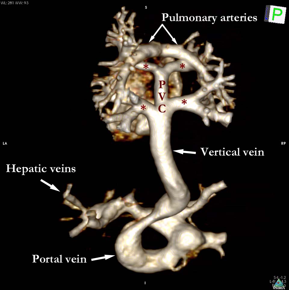
Modality: Cardiac MRI images (gadolinium-enhanced MR angiography) Orientation: Coronal plane (still & video images) Description: This neonatal patient has single ventricle physiology. The images are volume rendered 3D models derived from a magnetic resonance angiogram, captured during the first pass of contrast, which in this patient captures both the systemic and pulmonary venous phase. The prepared images begin in a coronal plane, and illustrate all the pulmonary veins connecting to a single, broad, descending vertical vein. This vertical vein is mildly narrowed within the liver, and connects to the hepatic portal system. (* - pulmonary veins, PVC - pulmonary venous confluence). Contributor: Marina Hughes, DPhil, MRCP, FRACP Institution: Centre for Cardiovascular Imaging, Great Ormond Street Hospital for Children NHS Trust, London, UK Image Label: MR040820-34c and MR040820-34d Source of Image: Centre for Cardiovascular Imaging, Great Ormond Street Hospital for Children NHS Trust, London, UK Image Certification: 10 May 2014
AWG Rating:
|
|
||
AWG Page Certification: 1 March 2014 (Recertification - 10 May 2014)
|
Copyright ipccc-awg.net All Rights Reserved. Frontpage-Templates.org |