|
||||||||
|
|
|||||||||
|
|||||||||
|
IPCCC: 06.02.18, 06.02.07, 04.01.26, 05.04.05 |
|||
|
AEPC Derived Term: |
Left ventricular inflow obstruction due to left superior caval vein (SVC) to coronary sinus (06.02.18) Mitral valvar stenosis: congenital (06.02.07) Left superior caval vein (SVC) persisting to coronary sinus to right-sided atrium (04.01.26) Atrial septal defect (ASD) within oval fossa (secundum): fenestrated (05.04.05) |
||
|
EACTS-STS Derived Term: |
Mitral valve disease, Mitral stenosis, Congenital, Supravalvar pathology, Mitral stenosis caused by left SVC to coronary sinus (06.02.18) Mitral valve disease, Mitral stenosis, Congenital (06.02.16) Systemic venous anomaly, SVC, Bilateral SVC, LSVC to CS (intact) to right-sided atrium (04.01.03, 04.01.26) ASD, Secundum, Fenestrated (05.04.05) |
||
|
Definition: pending
|
|
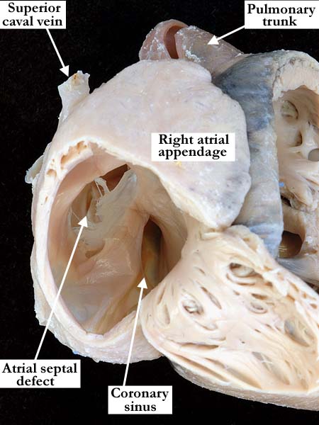
Modality: Anatomic specimen Orientation: Anterior oblique view Description: A window cut in the anterior lateral wall of the right atrium demonstrates the dilated coronary sinus and a fenestrated atrial septal defect. The right atrium and appendage are dilated. Contributor: Diane E. Spicer, BS Institution: The Congenital Heart Institute of Florida (CHIF) Image Label: A060218-95a Source of Image: Van Mierop Archive, University of Florida Image Certification: pending AWG Rating: pending
|
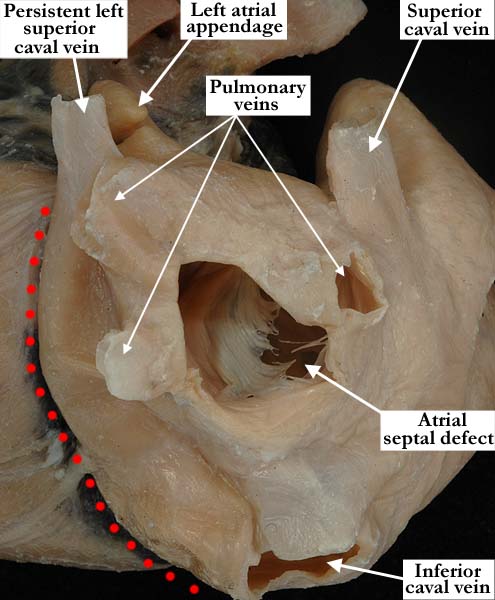
Modality: Anatomic specimen Orientation: Posterior oblique view Description: The red dots mark the external border of the large, persistent left superior caval vein as it extends over the posterior lateral aspect of the left atrium. The left atrial appendage is hypoplastic and the pulmonary veins are dilated. A fenestrated atrial septal defect is viewed through the window in the left atrium. Contributor: Diane E. Spicer, BS Institution: The Congenital Heart Institute of Florida (CHIF) Image Label: A060218-95b Source of Image: Van Mierop Archive, University of Florida Image Certification: pending AWG Rating: pending
|
|||
|
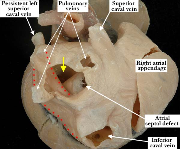
Modality: Anatomic specimen Orientation: Posterior view Description: The red dots demonstrate the large size of the persistent left superior caval vein as it extends to the coronary sinus. It partially obstructs the flow through the mitral valve and the pulmonary veins are dilated. There is mitral valve stenosis, the mitral valve marked by the yellow arrow. Contributor: Diane E. Spicer, BS Institution: The Congenital Heart Institute of Florida (CHIF) Image Label: A060218-95c Source of Image: Van Mierop Archive, University of Florida Image Certification: pending AWG Rating: pending
|
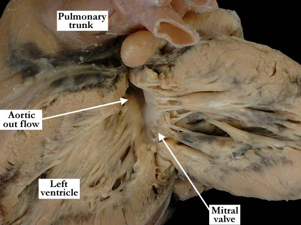
Modality: Anatomic specimen Orientation: Posterior view Description: The left ventricle has been opened in a clam-shell fashion, demonstrating the hypoplastic, stenotic mitral valve. The left ventricle is hypertrophied. Contributor: Diane E. Spicer, BS Institution: The Congenital Heart Institute of Florida (CHIF) Image Label: A060218-95d Source of Image: Van Mierop Archive, University of Florida Image Certification: pending AWG Rating: pending |
|||
AWG Page Certification: pending
|
Copyright ipccc-awg.net All Rights Reserved. Frontpage-Templates.org |