|
|||||
|
|
||||||
|
||||||
|
IPCCC: 04.04.03, 04.04.11, 01.04.04, 01.05.01, 07.02.00, 07.11.06 or
04.04.03, 04.04.11, 01.04.04,
01.03.00, 02.03.01, 02.06.02, 07.02.00, 01.05.01, 07.11.06 |
|||
|
AEPC Derived Term: |
Coronary sinus orifice atretic (04.04.03) Coronary sinus drainage cephalad to left superior caval vein (SVC) (04.04.11) Double inlet left ventricle (01.04.04) Discordant VA connections (TGA) (01.05.01) Right ventricular hypoplasia (07.02.00) Muscular VSD in outlet septum (07.11.06) |
||
|
EACTS-STS Derived Term: |
Systemic venous anomaly, SVC, CS ostial atresia or stenosis (CS draining cephalad via LSVC), CS ostial atresia (04.04.03, 04.04.11) Single ventricle, DILV, {SDD}, Subaortic RV outlet chamber with VSD (Bulboventricular foramen), (01.04.04, 01.03.00, 02.03.01, 02.06.02, 07.02.00) VA connection =Discordant VA connections (TGA) (01.05.01) VSD, Type 1 (Subarterial) (Supracristal) (Conal septal defect) (Infundibular), Conal muscular (07.11.06) |
||
|
Definition: pending
|
|
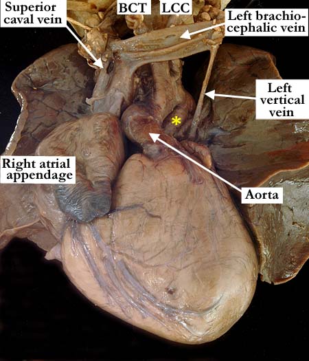
Modality: Anatomic specimen Orientation: Anterior view Description: This anterior view shows the aorta directly anterior to the pulmonary trunk in this heart with transposition of the great arteries (discordant ventriculo-arterial connections) and double inlet left ventricle. The aortic arch extends leftward and the brachiocephalic arteries branch from the arch in the usual fashion. The patent arterial duct (yellow asterisk) is left-sided. The superior caval vein and the left brachiocepalic vein are normal, with a small left veritical vein extending from the left brachiocephalic vein toward the posterior aspect of the heart. (BCT - brachiocephalic trunk, LCC - left common carotid artery) Contributor: Diane E. Spicer, BS Institution: The Congenital Heart Institute of Florida (CHIF) Image Label: A040403-82a Source of Image: The Congenital Heart Institute of Florida (CHIF) Image Certification: pending AWG Rating: pending
|
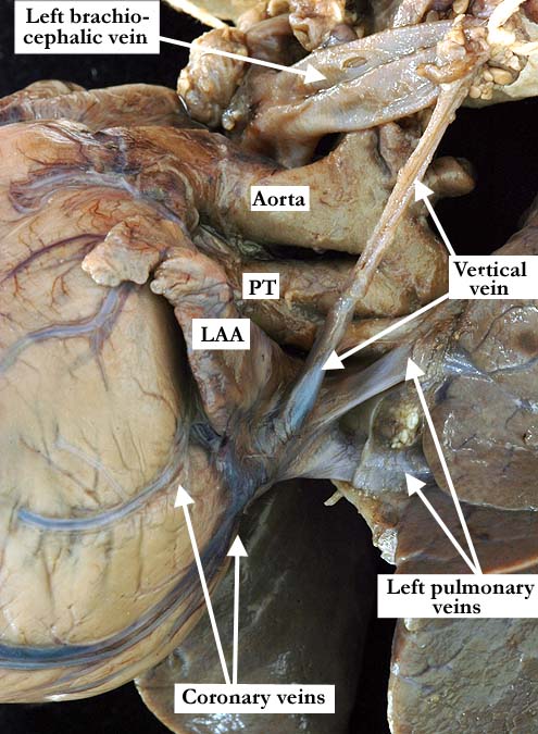
Modality: Anatomic specimen Orientation: Left, posterior, lateral view Description: In a left, posterior, lateral view the vertical vein shown in WA11-02a extends from the left brachiocephalic vein over the posterior aspect of the left atrium. This vertical vein is the mode of drainage for the cardiac venous blood secondary to atresia of the coronary sinus. Note the cardiac veins on the epicardial surface, draining directly into the vertical vein. (LAA - left atrial appendage, PT - pulmonary trunk) Contributor: Diane E. Spicer, BS Institution: The Congenital Heart Institute of Florida (CHIF) Image Label: A040403-82b Source of Image: The Congenital Heart Institute of Florida (CHIF) Image Certification: pending AWG Rating: pending |
|||
|
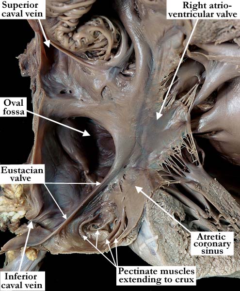
Modality: Anatomic specimen Orientation: Anterior lateral view Description: The anterior lateral view of this opened, dilated, right-sided, morphologically right atrium demonstrates the normal relationships of the superior and inferior caval veins to the right atrial chamber, along with a normal appearing Eustacian valve. There is no identifiable coronary sinus. The valve of the oval fossa is dilated and there is a small atrial septal defect along the superior rim. Contributor: Diane E. Spicer, BS Institution: The Congenital Heart Institute of Florida (CHIF) Image Label: A040403-82c Source of Image: The Congenital Heart Institute of Florida (CHIF) Image Certification: pending AWG Rating: pending
|
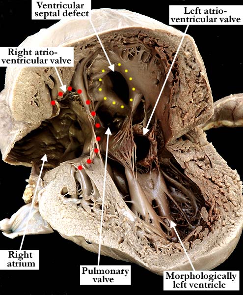
Modality: Anatomic specimen Orientation: Inferior apical view Description: This heart with double inlet left ventricle and transposition of the great arteries (discordant ventriculo-arterial connections) has been opened in a clam-shell fashion demonstrating the two atrioventricular valves connected to the dominant left ventricle. (Right atrioventricular valve outlined with red dots.) The interventricular septum has been lifted superiorly with the anterior portion of the heart, the ventricular septal defect marked with yellow dots. This defect is completely surrounded by muscle and is located between the apical and outlet portions of the ventricular septum. Contributor: Diane E. Spicer, BS Institution: The Congenital Heart Institute of Florida (CHIF) Image Label: A040403-82d Source of Image: The Congenital Heart Institute of Florida (CHIF) Image Certification: pending AWG Rating: pending
|
|||
|
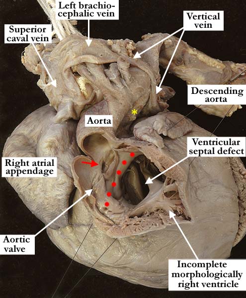
Modality: Anatomic specimen Orientation: Anterior view Description: This anterior, anatomic view demonstrates the typical antero-superior location of the incomplete, rudimentary right ventricle seen in those hearts with double inlet left ventricle. There are discordant ventriculo-arterial connections, the aorta exiting this rudimentary right ventricle above a subaortic infundibulum (red dots). The borders of the good-sized ventricular septal defect are entirely muscular. (red arrow-right coronary orifice, yellow asterisk-patent arterial duct) Contributor: Diane E. Spicer, BS Institution: The Congenital Heart Institute of Florida (CHIF) Image Label: A040403-82e Source of Image: The Congenital Heart Institute of Florida (CHIF) Image Certification: pending AWG Rating: pending
|
|
|||
AWG Page Certification: pending
|
Copyright ipccc-awg.net All Rights Reserved. Frontpage-Templates.org |