|
||||||||
|
|
|||||||||
|
|||||||||
|
IPCCC Code: 04.01.03, 04.01.26, 04.05.01 |
|||
|
AEPC Derived Term: |
Bilateral
superior caval veins (SVC) (04.01.03) |
||
|
EACTS-STS Derived Term: |
Systemic venous
anomaly, SVC, Bilateral SVC, LSVC to CS (intact) to right-sided atrium
(04.01.03, 04.01.26) |
||
|
ICD10 Derived Term: |
Other congenital malformations
of great veins (Q26.8) |
||
|
Definition: NA |
|
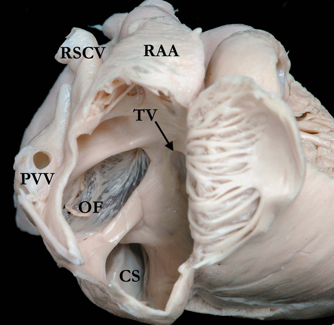
Modality: Anatomic specimen Orientation: Anterior right atrial view Description: The anterior wall of this morphologically right atrium has been reflected to show the atrial septum in anatomic position. The atrial septum, including the oval fossa (OF) is intact and the opening of the coronary sinus (CS) into the right atrium is dilated. This dilatation is secondary to a persistent left superior caval vein draining to it. (RSCV-superior caval vein, RAA-right atrial appendage, TV-tricuspid valve, PVV-pulmonary vein). Contributor: Diane Spicer, BS Institution: The Congenital Heart Institute of Florida (CHIF) Image Label: A040103-33a Source of Image: Van Mierop Archive, University of Florida, Gainesville, Florida Image Certification: 10 May 2014
AWG Rating:
|
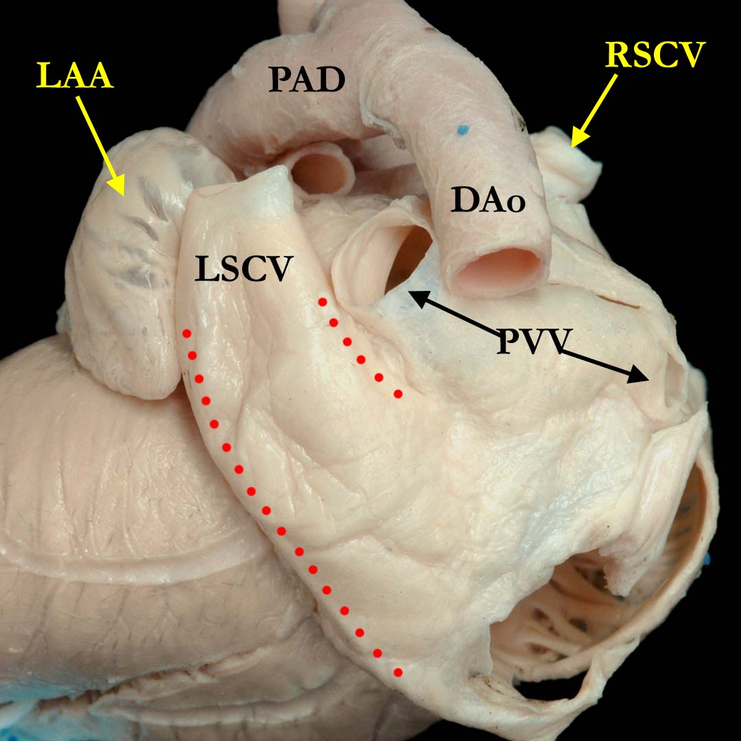
Modality: Anatomic specimen Orientation: Posterior view Description: This posterior view of the heart pictured in image one demonstrates the persistent left superior caval vein (LSCV) extending into the coronary sinus (CS) (red dots) over the posterior aspect of the left atrium. The pulmonary veins (PVV) drain to the left atrium in the usual fashion, just to the right of the persistent left superior caval vein. (PAD-patent arterial duct, DAo-descending aorta, LAA-left atrial appendage, RSCV-right superior caval vein). Contributor: Diane Spicer, BS Institution: The Congenital Heart Institute of Florida (CHIF) Image Label: A040103-33b Source of Image: Van Mierop Archive, University of Florida, Gainesville, Florida Image Certification: 10 May 2014
AWG Rating:
|
|||
|
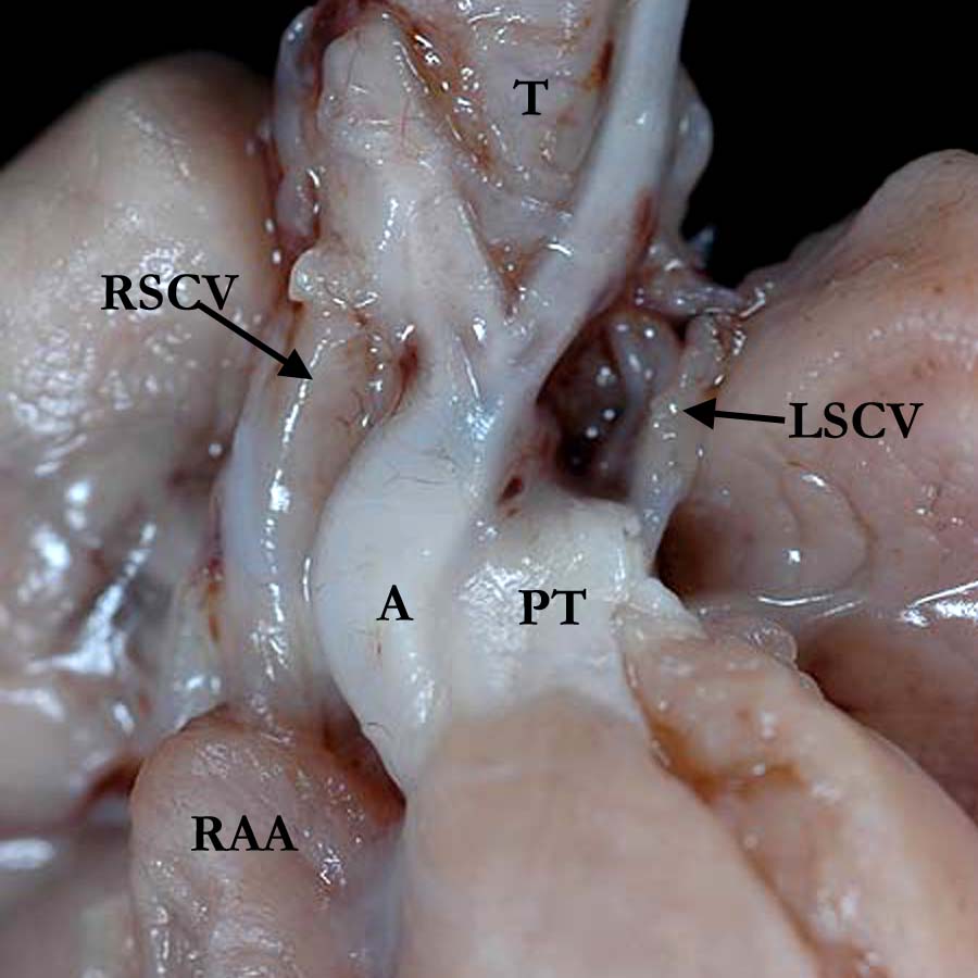
Modality: Anatomic specimen Orientation: Anterior view Description: The anterior, anatomic view of a different heart in a fetus at 16 weeks gestation demonstrates absence of the brachiocephalic (innominate) vein with an otherwise normal superior caval vein (RSCV) on the right and a persistent left superior caval vein (LSCV) on the left. The position of the aorta (A) is slightly anterior and to the right. The great vessels branch normally from the aortic arch. (PT- pulmonary trunk, RAA-right atrial appendage, T-trachea). Contributor: Diane Spicer, BS Institution: The Congenital Heart Institute of Florida (CHIF) Image Label: A040103-33c Source of Image: The Congenital Heart Institute of Florida (CHIF) Image Certification: 10 May 2014
AWG Rating:
|
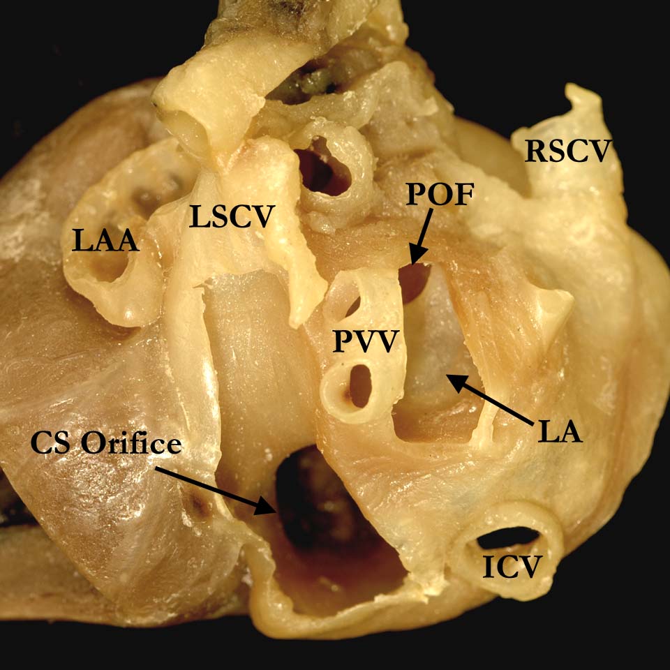
Modality: Anatomic specimen Orientation: Posterior view
Description: This image is from another
heart that is viewed posteriorly in anatomic position and is parafinized.
The walls of the left atrium
(LA) and the persistent left superior caval
vein (LSCV) have been dissected away to illustrate that the two structures
are totally separate from one another. The persistent left superior caval
vein connects to the coronary sinus which extends along the posterior wall
of the left atrium to the crux of the heart. The opening orifice of the
coronary sinus is dilated. The patent oval foramen
(POF) is visualized
within the left atrial cavity and the pulmonary veins (PVV) drain to the left
atrium in the usual fashion. (LAA-left atrial appendage, RSCV-superior caval
vein,
ICV-inferior caval vein, PVV-pulmonary veins) Institution: The Congenital Heart Institute of Florida (CHIF) Image Label: A040103-33d Source of Image: Children's Hospital Colorado, Aurora, Colorado Image Certification: 10 May 2014
AWG Rating:
|
AWG Certification: 10 May 2014
|
Copyright ipccc-awg.net All Rights Reserved. Frontpage-Templates.org |