|
|||||
|
|
||||||
|
||||||
|
IPCCC: 01.01.25, 09.10.45 |
|||
|
AEPC Derived Term: |
Pulmonary atresia + VSD + systemic-to-pulmonary collateral artery(ies) (MAPCA(s)), (01.01.25) Confluent pulmonary arteries (09.10.45) |
||
|
EACTS-STS Derived Term: |
Pulmonary atresia-VSD-MAPCA(s) (Pseudotruncus), Type B (Native PA present and MAPCA(s) present), Confluent RPA and LPA, (01.01.25, 09.10.45) |
||
|
Definition: pending
|
|
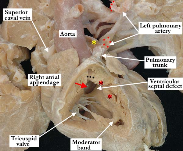
Modality: Anatomic specimen Orientation: Anterior view Description: In this anterior, anatomic view, the right ventricle has been windowed to view the components of the right ventricle. The tricuspid valve guards the inlet and the right ventricle is hypertrophied. There is anterior deviation (black dots) of the outlet septum as well as hypertrophy of the septo-parietal trabeculations (red asterisks) causing significant narrowing of the subpulmonary infundibulum. Note the discrepant size between the aorta and the pulmonary artery, the aorta anterior and to the right of its usual position. There is a perimembranous ventricular septal defect with the aortic valve (red arrow) visible at its roof. The pulmonary artery is small and there is pulmonary atresia. The left main pulmonary artery was cut during dissection and the opposing edges are marked with red dots. The right pulmonary artery (yellow asterisk) branches normally from the pulmonary trunk and extends posterior to the aorta. Contributor: Diane E. Spicer, BS Institution: The Congenital Heart Institute of Florida (CHIF) Image Label: A010125-86a Image Source: Van Mierop Archive, University of Florida Image Certification: pending AWG Rating: pending
|
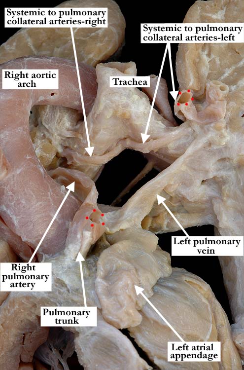
Modality: Anatomic specimen Orientation: Superior, left lateral view Description: In this superior, left, lateral view this heart with tetralogy of Fallot and pulmonary atresia demonstrates a large aorta with a right aortic arch and a small pulmonary trunk giving rise to small right and left pulmonary arteries. (Note: the left pulmonary artery was cut during dissection and the opposing ends are marked with red dots). Several systemic to pulmonary collateral arteries are extending from the descending aorta to both the right and left lungs, on the left, a few arteries are connecting directly to the left main pulmonary artery. Contributor: Diane E. Spicer, BS Institution: The Congenital Heart Institute of Florida (CHIF) Image Label: A010125-86b Image Source: Van Mierop Archive, University of Florida Image Certification: pending AWG Rating: pending |
|||
|
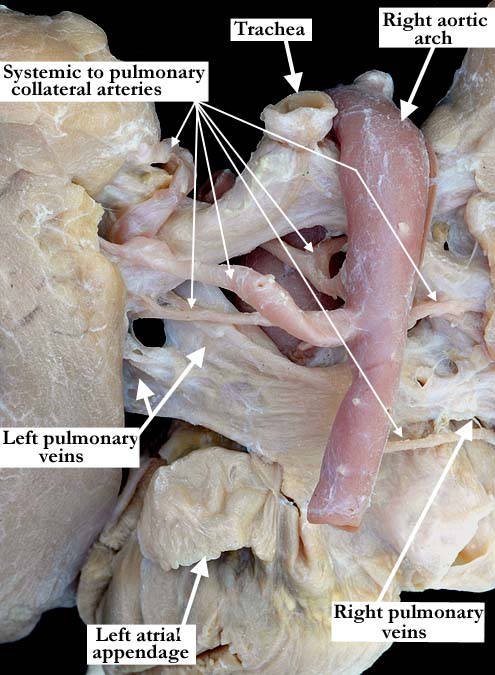
Modality: Anatomic specimen Orientation: Posterior, left, lateral view Description: A posterior, left lateral view of the right aortic arch and descending aorta, nicely demonstrates multiple systemic to pulmonary collateral arteries. Contributor: Diane E. Spicer, BS Institution: The Congenital Heart Institute of Florida (CHIF) Image Label: A010125-86c Image Source: Van Mierop Archive, University of Florida Image Certification: pending AWG Rating: pending
|
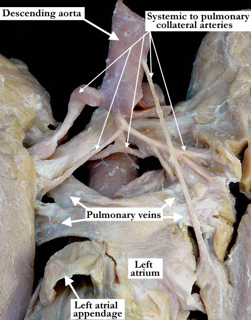
Modality: Anatomic specimen Orientation: Posterior view Description: In this posterior view, the descending aorta has been lifted superiorly to demonstrate the multiple, systemic to pulmonary collateral arteries, several of which arise from the anterior aspect of the aorta. Contributor: Diane E. Spicer, BS Institution: The Congenital Heart Institute of Florida (CHIF) Image Label: A010125-86d Image Source: Van Mierop Archive, University of Florida Image Certification: pending AWG Rating: pending
|
AWG Page Certification: pending
|
Copyright ipccc-awg.net All Rights Reserved. Frontpage-Templates.org |