|
||||||||
|
|
|||||||||
|
|||||||||
|
IPCCC: 04.08.01, Q1.01.51 |
|||
|
AEPC Derived Term: |
Pulmonary venous stenosis - congenital - all pulmonary veins (04.08.01, Q1.01.51) |
||
|
EACTS-STS Derived Term: |
Pulmonary venous stenosis, Congenital (04.08.01) Pulmonary veins-modifier for vein(s) involved, All pulmonary veins (Q1.01.51) |
||
|
ICD10 Derived Term: |
Other congenital malformations of great veins (Q26.8) |
||
|
Definition: pending
|
|
Modality: Anatomic specimen Orientation: Inferior view Description: The heart has been lifted cephalad. The inferior or diaphragmatic aspect of the right atrium shows the inferior caval vein and just posterior to that are the right and left pulmonary veins as they join the left atrium. The right pulmonary veins join the left atrium via a short, stenotic vein. There are two right lower pulmonary veins, one of which is small and the other, large and dilated. The dilated right lower pulmonary vein crosses the midline to drain into the left atrium along with the left pulmonary veins. The entrance of the left pulmonary veins into the left atrium is also markedly stenotic. This specimen also has an abnormal tricuspid valve, illustrated in its companion web page. Contributor: Diane E. Spicer, BS Institution: The Congenital Heart Institute of Florida (CHIF) Image Label: A040801-101a Source of Image: The Congenital Heart Institute of Florida (CHIF) Image Certification: pending AWG Rating: pending
|
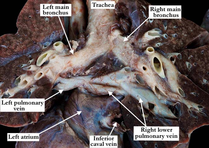
Modality: Anatomic specimen Orientation: Posterior view Description: This posterior view shows the cut surfaces of the lungs and morphologically normal right and left main bronchuses. The right lower pulmonary vein is markedly dilated as it extends into the parenchyma and toward the periphery of the lung. This is secondary to the severe stenosis as the veins enter the left atrium. Note how the right lower pulmonary vein crosses the midline to drain into the left atrium. Contributor: Diane E. Spicer, BS Institution: The Congenital Heart Institute of Florida (CHIF) Image Label: A040801-101b Source of Image: The Congenital Heart Institute of Florida (CHIF) Image Certification: pending AWG Rating: pending |
|||
|
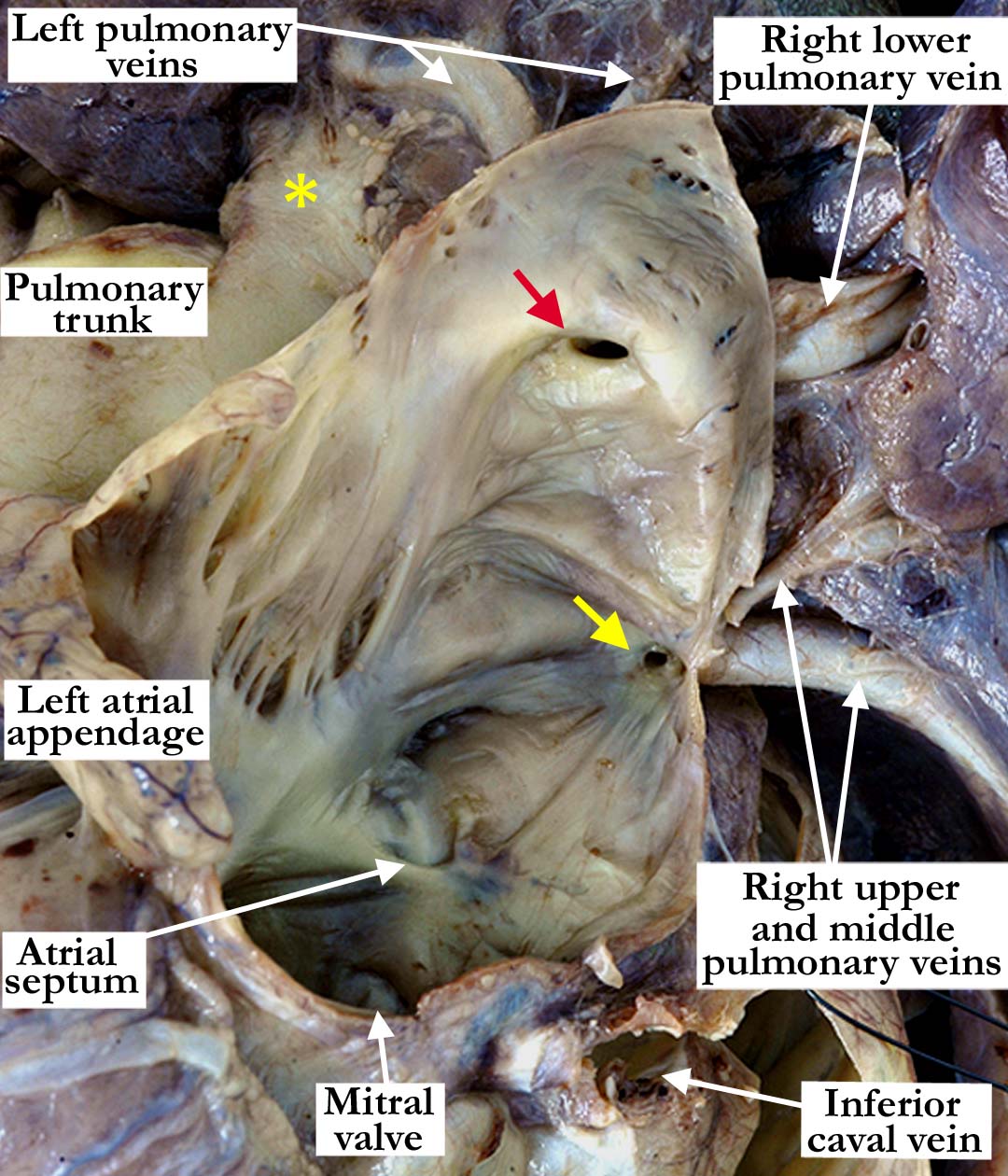
Modality: Anatomic specimen Orientation: Posterior view Description: The left atrium has been opened and the lungs have been lifted in a cephalic direction . The left pulmonary veins and the right lower pulmonary vein drain into the left atrium via a single, stenotic opening (red arrow). The right upper and middle pulmonary veins drain via an even more stenotic opening (yellow). The atrial septum is intact. (yellow asterisk left pulmonary artery). Institution: The Congenital Heart Institute of Florida (CHIF) Image Label: A040801-101c Source of Image: The Congenital Heart Institute of Florida (CHIF) Image Certification: pending AWG Rating: pending
|
||||
AWG Page Certification: pending
|
Copyright ipccc-awg.net All Rights Reserved. Frontpage-Templates.org |