|
(click image to
view original size) |
|
Derived Terms: |
|
|
AEPC: |
Normal heart (01.01.00) |
| |
Normal position-orientation of heart (02.01.00) |
| |
|
|
EACTS-STS: |
Normal heart (01.01.00) |
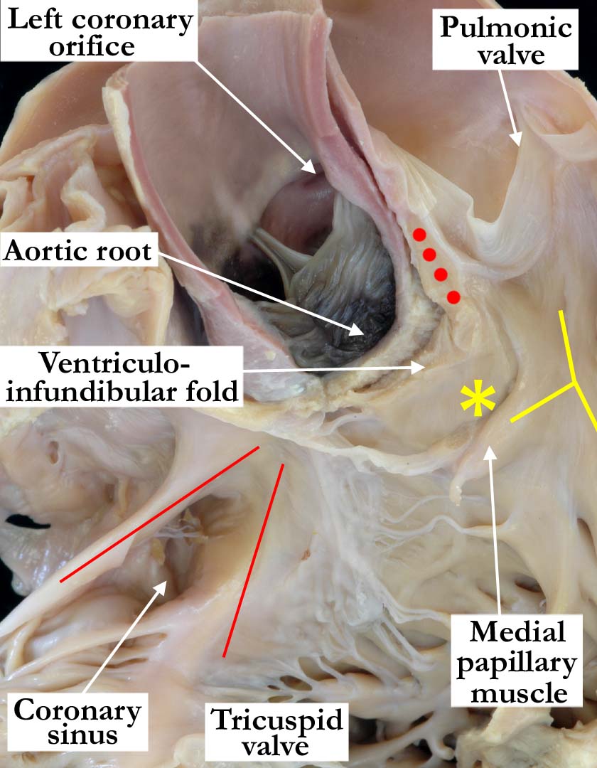
Modality: Anatomic specimen
Orientation: Anterior view
Description: In this close up view, the
anterior free wall of the right ventricle, the anterior pulmonary trunk and
anterior-most wall of the aorta have been removed. All three leaflets of the
aortic valve remain intact within the aortic root. The free standing
muscular sleeve (red dots), or subpulmonary infundibulum, supports the
leaflets of the pulmonary valve. It is an integral part of the
supraventricular crest, although the majority of the crest is formed by the
ventriculo-infundibular fold, or inner heart curvature, which has been cut
away produce this image. A small area, marked with the yellow asterisk,
represents the outlet component of the muscular septum, at the point where
the crest joins the septomarginal trabeculation (yellow Y). There are no
anatomic boundaries, however, showing where this component begins or ends.
Note the triangle of Koch (red lines), delineated by the tendon of Todaro
and the hinge of the septal leaflet of the tricuspid valve.
Contributor: Diane
E. Spicer, BS
Institution: The Congenital Heart
Institute of Florida (CHIF)
Image Label: A010100-148a
Source of Image: The Congenital Heart
Institute of Florida (CHIF)
Image Certification: pending
AWG Rating: pending
|
|
