IPCCC: 01.01.00, 02.01.00
|
|||||||||
|
|
|
IPCCC: 01.01.00, 02.01.00 |
|
(click image to view original size) |
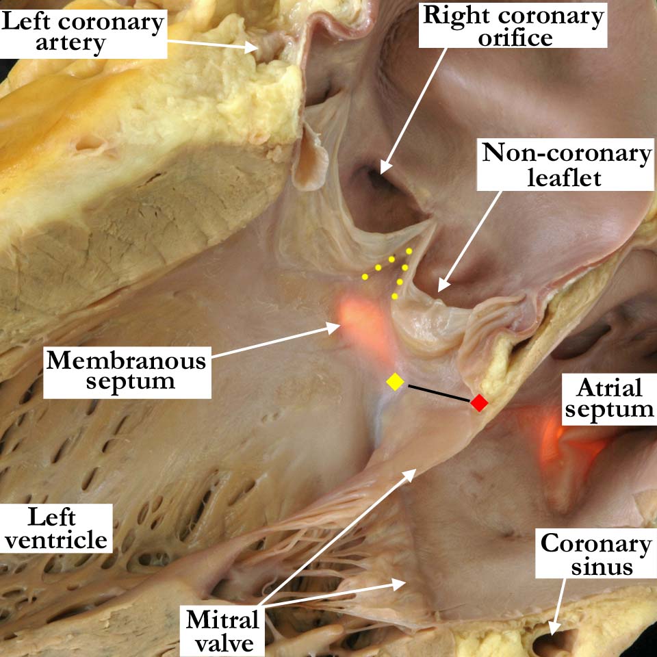
Modality: Anatomic specimen Orientation: Posterior left ventricular view
Description: The left ventricular aspect
of the same heart shown in image
A010100-134a
has a light placed on the right side of the membranous septumThe membranous
septum is seen to be continuous with the interleaflet fibrous triangle
(yellow dots) positioned beneath the zone of apposition between the
non-coronary and right coronary leaflets of the aortic valve. The fibrous
trigones (yellow and red diamonds)_ are the thickened areas at the ends of
the zone of fibrous continuity between the leaflets of the aortic and mitral
valves (black line). The so-called central fibrous body, the strongest part
of the fibrous skeleton of the heart, is formed by the union of the right
fibrous trigone and the membranous septum. Institution: The Congenital Heart Institute of Florida (CHIF) Image Label: A010100-135a Source of Image: The Congenital Heart Institute of Florida (CHIF) Image Certification: pending AWG Rating: pending |
|||||||||||
AWG Page Certification: pending
|
Copyright ipccc-awg.net All Rights Reserved. Frontpage-Templates.org |