|
||||||||
|
|
|||||||||
|
|||||||||
|
IPCCC: 06.02.09, 06.02.05, Q1.27.75, 05.02.04, 07.11.02 |
|||
|
AEPC Derived Term: |
Straddling mitral valve (06.02.09) Overriding mitral valve - override of AV valve 50-90% (06.02.05, Q1.27.75) Left atrial appendage (right) juxtaposition (05.02.04) Muscular VSD in inlet septum (07.11.02) |
||
|
EACTS-STS Derived Term: |
Mitral valve disease, Mitral valve pathology, Straddling mitral valve (06.02.09) Mitral valve disease, Mitral valve pathology, Overriding mitral valve, Override of mitral valve 50-90% (06.02.05, Q1.27.75) Atrial abnormality, Juxtaposition of the atrial appendages, LA appendage (right) juxtaposition (05.02.04) VSD, Type 4 (Muscular), Inlet (Posterior) (07.11.02) |
||
|
Definition: pending
|
|
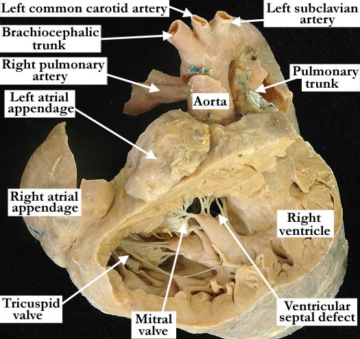
Modality: Anatomic specimen Orientation: Anterior view Description: The free wall of the right ventricle is removed to demonstrate the straddling and overriding mitral valve. There are cordal attachments to a right ventricular papillary muscle as well as to the left ventricular papillary muscles (shown in fig. 3). The tricuspid valve enters the right ventricle inferior to the mitral valve. The right ventricle is hypertrophied and there is a pulmonary artery band in place. There are concordant ventriculo-arterial connections and the atrial appendages are juxtaposed to the right of the arterial pedicle. Contributor: Diane E. Spicer, BS Institution: The Congenital Heart Institute of Florida (CHIF) Image Label: A060209-98a Source of Image: Van Mierop Archive, University of Florida Image Certification: pending AWG Rating: pending
|
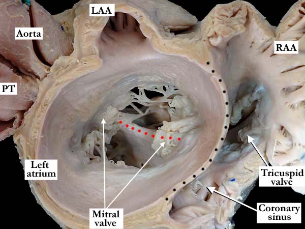
Modality: Anatomic specimen Orientation: Short axis at the base of the heart Description: This close-up, short axis view at the base of the heart clearly demonstrates the atrioventricular septal malalignment along with the straddling and overriding mitral valve. The ventricular septum (red dots) lies perpendicular to the atrial septum (black dots), which is dilated toward the right atrium. Approximately 50 % of the mitral valve circumference lies within the right ventricle. The aorta lies slightly more to the right of the pulmonary trunk than usual. The atrial appendages are juxtaposed to the right of the vascular pedicle and there is marked dilatation of the left atrium. (LAA-left atrial appendage, RAA-right atrial appendage) Contributor: Diane E. Spicer, BS Institution: The Congenital Heart Institute of Florida (CHIF) Image Label: A060209-98b Source of Image: Van Mierop Archive, University of Florida Image Certification: pending AWG Rating: pending |
|||
|
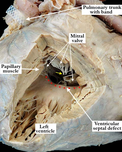
Modality: Anatomic specimen Orientation: Posterior view Description: Through the window cut in the free wall of the left ventricle the ventricular septal defect is easily appreciated. There is left ventricular hypertrophy. The tendinous cords of the mitral valve attach to papillary muscles within both the right and left ventricles. One left ventricular papillary muscle is left intact and the small yellow arrow illustrates the cordal attachments that extended to the resected papillary muscle. (red dots-crest of the ventricular septum) Contributor: Diane E. Spicer, BS Institution: The Congenital Heart Institute of Florida (CHIF) Image Label: A060209-98c Source of Image: Van Mierop Archive, University of Florida Image Certification: pending AWG Rating: pending
|
||||
AWG Page Certification: pending
|
Copyright ipccc-awg.net All Rights Reserved. Frontpage-Templates.org |